ADVERTISEMENT

The winners of the 51st annual Nikon Small World Photomicrography Competition have been announced, showcasing some of the most stunning and detailed images of the microscopic world. From insects to plant structures, these photographs reveal the beauty and complexity of tiny subjects we often overlook in our daily lives. The competition, run by Nikon Instruments, has been celebrating excellence in microscopy and digital imaging for over five decades, inspiring scientists and artists alike.

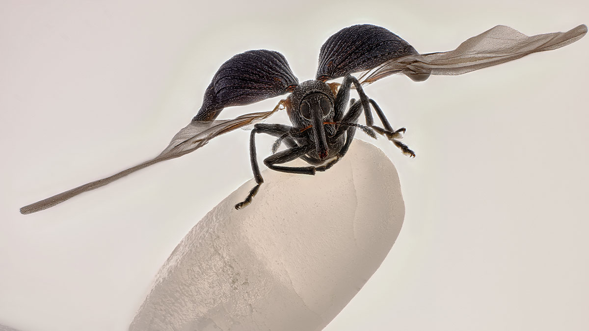
This year’s top prize went to China’s Zhang You for his incredible image of a rice weevil perched on a grain of rice, wings fully extended. Using a combination of photography skills, careful lighting, and focus stacking, You captured a moment that blends scientific detail with artistic flair. His work reminds us that even the smallest creatures can be fascinating and beautiful when seen up close.

More info: nikonsmallworld.com | Instagram | Facebook | x.com

#1

3rd Place: John-Oliver Dum, Medienbunker Produktion, Bendorf, Rheinland Pfalz, Germany

Yellow spherical pollen grains clinging to water droplets on a delicate spider web in a mind-blowing microscopic image.

"Pollen in a garden spider web."

© John-Oliver Dum | Nikon Small World Report

Zhang You didn’t just win first place; he also earned 15th place with a second image showing a Geometer moth laying eggs. A member of the Entomological Society of China, You has spent years studying insects and teaching others about them. He says the key to a great microscopic photo is a mix of science and art, from understanding the subject’s behavior to mastering lighting. “It pays to dive deep into entomology: understanding insects’ behaviors and mastering lighting,” You said. “A standout work blends artistry with scientific rigor, capturing the very essence, energy, and spirit of these creatures.”

RELATED:
    #2

    Honorable Mention: Gregory B. Murray, Pritchard, British Columbia, Canada

    Close-up microscopic image showing detailed crystalline structures in the 2025 Nikon Small World photomicrography contest.

    "Frost on a wooden railing."

    © Gregory B. Murray | Nikon Small World Report

    sbj
    Community Member
    1 month ago Created by potrace 1.15, written by Peter Selinger 2001-2017

    Amazing! I'm not going to brush my hand along a rail, I'm just going to let it lie

    ADVERTISEMENT
    #3

    Image Of Distinction: Ye Fei Zhang, Jiang Yin, Jiang Su, China

    Close-up microscopic image of red, textured structures on a rough surface from the 2025 Nikon Small World photomicrography contest

    "Butterfly (Artopoetes pryeri) eggs."

    © Ye fei Zhang | Nikon Small World Report

    C B Jones
    Community Member
    1 month ago Created by potrace 1.15, written by Peter Selinger 2001-2017

    These look like little crochet hats, they're so darn cute!

    View more comments

    “Zhang You’s work demonstrates the remarkable power of microscopy to reveal new perspectives on the world around us,” said Eric Flem, Senior Manager, Communications and CRM at Nikon Instruments. “What makes this year even more extraordinary is that it was his very first time entering the competition, and he not only captured first place, but also placed another image in the top 20. His achievement highlights the spirit of Nikon Small World: inspiring wonder, making scientific understanding accessible to all, and celebrating the artistry of the microscopic realm.”

    #4

    Image Of Distinction: Marek Miś, Marek Miś Photography, Suwalki, Podlaskie, Poland

    Colorful microscopic crystals with sharp edges captured in a vibrant Nikon Small World photomicrography contest image.

    "Crystallized soy sauce."

    © Marek Miś | Nikon Small World Report

    #5

    Image Of Distinction: Dr. David Maitland, Art Of Science, St. Andrews, Fife, United Kingdom

    Close-up microscopic image of plant cells with vibrant colors, featured in the 2025 Nikon Small World photomicrography contest.

    "Vascular bundles in a bamboo leaf (Phyllostachys sp.)."

    © Dr. David Maitland | Nikon Small World Report

    ADVERTISEMENT

    The second-place winner, Dr. Jan Rosenboom from Germany, captured stunning spheres of Volvox algae in a drop of water, while third place went to John-Oliver Dum, also from Germany, for an intricate photo of pollen caught in a garden spider’s web. Both images reveal patterns and details that most people would never notice in everyday life.

    #6

    2nd Place: Dr. Jan Rosenboom, Rostock, Mecklenburg Vorpommern, Germany

    Close-up of a glowing green microscopic droplet with bubble-like structures in the 2025 Nikon Small World photomicrography contest.

    "Colonial algae (Volvox) spheres in a drop of water."

    © Dr. Jan Rosenboom | Nikon Small World Report

    #7

    5th Place: Dr. Igor Siwanowicz, Howard Hughes Medical Institute (Hhmi), Janelia Research Campus, Ashburn, Virginia, USA

    Colorful microscopic image showcasing intricate cellular structures from the 2025 Nikon Small World photomicrography contest.

    "Spores (blue/purple structures) of a small tropical fern (Ceratopteris richardii)."

    © Dr. Igor Siwanowicz | Nikon Small World Report

    Many of the top images use a technique called “image stacking,” where multiple photos are combined to create one sharp, detailed picture. Others rely on confocal or fluorescence microscopy to highlight structures inside cells or tiny organisms, letting viewers see hidden shapes and colors that would otherwise be invisible.

    ADVERTISEMENT
    #8

    11th Place: Marek Miś, Marek Miś Photography, Suwalki, Podlaskie, Poland

    Close-up microscopic image showing colorful detailed structures, featured in the 2025 Nikon Small World photomicrography contest.

    "Sunflower trichomes (hair-like plant outgrowths)."

    © Marek Miś | Nikon Small World Report

    ADVERTISEMENT
    #9

    Honorable Mention: Michael Parra Puentes, Bogotá, Cundinamarca, Colombia

    Close-up microscopic image showing detailed yellow hairs on an insect claw from the 2025 Nikon Small World photomicrography contest.

    "Thoracic and cephalic horn of a male beetle (Golofa porteri)."

    © Michael Parra Puentes | Nikon Small World Report

    In total, the competition recognized 71 images from thousands of entries worldwide. From crystallized soy sauce to mouse neurons, the photos show the incredible variety of life under the microscope—and how curiosity and patience can turn even the tiniest subjects into breathtaking works of art.

    #10

    13th Place: Henri Koskinen, Helsinki University, Helsinki, Uudenmaan Lääni, Finland

    Microscopic image showing highly detailed red fungal structures captured in the 2025 Nikon Small World photomicrography contest.

    "Slime mold (Arcyria major) releasing spores."

    © Henri Koskinen | Nikon Small World Report

    S Bow
    Community Member
    1 month ago Created by potrace 1.15, written by Peter Selinger 2001-2017

    Slime molds are fascinating, they can physically move to a better location if their current one isn't optimal.

    View more comments
    ADVERTISEMENT
    #11

    14th Place: Manfred Heising, Lwl Museum Of Natural History Münster, Münster, Northrhine-Westphalia, Germany

    Microscopic image showing intricate yellow mineral filaments inside a crystal from the 2025 Nikon Small World contest.

    "Quartz with biotic goethite filaments."

    © Manfred Heising | Nikon Small World Report

    Gebidozo
    Community Member
    1 month ago Created by potrace 1.15, written by Peter Selinger 2001-2017

    Ahhh… the magical Crystal of Power! Now I can finally control this world! Beware, puny humans! Mwaaaa-hhhahahahahaa!!..

    View more comments
    #12

    Image Of Distinction: Dr. Gonzalo Quiroga Artigas, Crbm-Cnrs, Montpellier, Herault, France

    Microscopic image of a transparent tardigrade highlighting internal structures in the 2025 Nikon Small World contest.

    "Tardigrade."

    © Dr. Gonzalo Quiroga Artigas | Nikon Small World Report

    #13

    Honorable Mention: Michael Robert Peres, Rochester Institute Of Technology, School Of Photographic Arts And Sciences, Rochester, New York, USA

    Close-up microscopic image of a detailed snowflake from the 2025 Nikon Small World photomicrography contest.

    "Melting snowflake."

    © Michael Robert Peres | Nikon Small World Report

    Gebidozo
    Community Member
    1 month ago Created by potrace 1.15, written by Peter Selinger 2001-2017

    An encoded message from a technologically advanced alien civilization!

    #14

    Image Of Distinction: Daniel Evrard, Aywaille, Liege, Belgium

    Close-up microscopic image revealing intricate patterns and textures from the 2025 Nikon Small World photomicrography contest.

    "Androconial (pheromone producing) area of a butterfly (Colias) wing."

    © Daniel Evrard | Nikon Small World Report

    setsuriseikou
    Community Member
    1 month ago Created by potrace 1.15, written by Peter Selinger 2001-2017

    Wait, those are on their wings? Not body?

    #15

    Image Of Distinction: Dr. Francisco Lázaro-Diéguez, Albert Einstein College Of Medicine, Bronx, New York, USA

    Microscopic image showing intricate swirling patterns and vibrant colors from the Nikon Small World photomicrography contest

    "Dedifferentiated liver cell."

    © Dr. Francisco Lázaro-Diéguez | Nikon Small World Report

    #16

    Image Of Distinction: Jianguo Mao, Shanghai, Shanghai, China

    Microscopic image of a colorful water flea showing intricate details in the 2025 Nikon Small World photomicrography contest.

    "Pregnant water flea (Daphnia)."

    © Jianguo Mao | Nikon Small World Report

    #17

    1st Place: Zhang You, Kunming, Yunnan, China

    Close-up microscopic image of an insect with detailed wings and body from the 2025 Nikon Small World contest.

    "Rice weevil (Sitophilus oryzae) on a grain of rice."

    © Zhang You | Nikon Small World Report

    sbj
    Community Member
    1 month ago Created by potrace 1.15, written by Peter Selinger 2001-2017

    Always disliked the pesky little things after once getting in my rice but now I see a close up and can sort of forgive them

    View more comments
    #18

    Image Of Distinction: Karl Deckart, Eckental, Bavaria, Germany

    Colorful abstract pattern captured in a mind-blowing microscopic image from the 2025 Nikon Small World contest.

    "Recrystallization of phenyl imidazol."

    © Karl Deckart | Nikon Small World Report

    #19

    Image Of Distinction: Dr. Michael Weber, Berlin Institute Of Health At Charité, Department Of Human Genetics, Berlin, Germany

    Microscopic image showcasing intricate glowing cellular structures captured in the Nikon Small World photomicrography contest.

    "Blood vessels in the limb of an embryonic mouse."

    © Dr. Michael Weber | Nikon Small World Report

    #20

    Honorable Mention: Dr. Bruno Cisterna And Dr. Eric Vitriol, Medical College Of Georgia At Augusta University, Department Of Neuroscience & Regenerative Medicine, Augusta, Georgia, USA

    Fluorescent microscopic image showcasing intricate neural networks with vibrant purple and blue structures from Nikon Small World contest.

    "Human neurons reprogrammed from skin cells."

    © Dr. Bruno Cisterna and Dr. Eric Vitriol | Nikon Small World Report

    Multa Nocte
    Community Member
    1 month ago Created by potrace 1.15, written by Peter Selinger 2001-2017

    Cool! I used to be on the faculty at MCG Augusta!

    View more comments
    #21

    Image Of Distinction: Jonathan Muyal, Paris, France

    Colorful abstract pattern of microscopic crystals captured in a mind-blowing 2025 Nikon Small World photomicrography image

    "Iridescent rutile (mineral) needles in a Burmese ruby."

    © Jonathan Muyal | Nikon Small World Report

    Gebidozo
    Community Member
    1 month ago (edited) Created by potrace 1.15, written by Peter Selinger 2001-2017

    The beginning of a trailer for a new cutting-edge cyberpunk movie.

    #22

    Image Of Distinction: Marek Miś, Marek Miś Photography, Suwalki, Podlaskie, Poland

    Microscopic image from the 2025 Nikon Small World contest showing colorful bubbles with a heart shape under a microscope.

    "Air bubbles in melted polyvinyl alcohol."

    © Marek Miś | Nikon Small World Report

    #23

    Image Of Distinction: Dr. Stephen De Lisle, Karlstad University, Department Of Environmental And Life Sciences, Karlstad, Värmland, Sweden

    Microscopic image of patterned seeds with green and brown hues, showcasing detail from the 2025 Nikon Small World photomicrography contest.

    "Lily flower pollen (autofluorescence)."

    © Dr. Stephen de Lisle | Nikon Small World Report

    #24

    Image Of Distinction: Walter Ferrari, Walter Ferrari Macro, Rio Cuarto, Cordoba, Argentina

    Close-up microscopic image of insect eggs captured in the 2025 Nikon Small World photomicrography contest.

    "True bug (Hemipteran) eggs on a leaf."

    © Walter Ferrari | Nikon Small World Report

    #25

    16th Place: Rogelio Moreno, Panama, Panama

    Close-up microscopic image showcasing vibrant green and blue structures from the 2025 Nikon Small World photomicrography contest.

    "Spore sacs (sporangia) of a fern."

    © Rogelio Moreno | Nikon Small World Report

    Sonder Toffee
    Community Member
    1 month ago Created by potrace 1.15, written by Peter Selinger 2001-2017

    Ferns are funkier than I ever imagined

    #26

    Honorable Mention: Jiri Cerny, Institute Of Molecular Genetics Of The Czech Academy Of Sciences, Light Microscopy Core Facility, Prague, Czech Republic

    Close-up microscopic image of a spider showcasing intricate details in the 2025 Nikon Small World photomicrography contest.

    "Jumping spider."

    © Jiri Cerny | Nikon Small World Report

    #27

    Image Of Distinction: Joe Mckellar, Cnrs, The Institue Of Molecular Genetics Of Montpellier (Igmm), Montpellier, Hérault, France

    Microscopic image showcasing intricate cellular structures captured in the 2025 Nikon Small World photomicrography contest.

    "Giant human hepatic cancer cell surrounded by smaller cells."

    © Joe Mckellar | Nikon Small World Report

    #28

    Image Of Distinction: Dr. Mette Handberg-Thorsager, Alexandre Alié And Lisa Maria Ulbrich, Georg-August-University Göttingen, Department Of Multiscale Biology, Göttingen, Niedersachsen, Germany

    Microscopic image of a detailed translucent organism with red and white structures from the Nikon Small World contest.

    "Oozoid of a sea squirt (Thalia democratica)."

    © Dr. Mette Handberg-Thorsager, Alexandre Alié and Lisa Maria Ulbrich | Nikon Small World Report

    Gebidozo
    Community Member
    1 month ago Created by potrace 1.15, written by Peter Selinger 2001-2017

    Thalia DEMOCRATICA? Is that… a great country?😳

    View more comments
    #29

    Image Of Distinction: Dr. Noah Bressman, Salisbury University, Department Of Biology, Salisbury, Maryland, USA

    Microscopic image of a fish skeleton with vibrant purple and yellow hues from the 2025 Nikon Small World photomicrography contest.

    "Histologically-stained harvestfish/star butterfish (Peprilus paru)."

    © Dr. Noah Bressman | Nikon Small World Report

    pelemele
    Community Member
    1 month ago Created by potrace 1.15, written by Peter Selinger 2001-2017

    Amazingly, it's as if he had three spinal columns.

    View more comments
    #30

    Image Of Distinction: Solvin Zankl, Kiel, Schleswig-Holstein , Germany

    Close-up microscopic image showcasing intricate blue and purple details from the 2025 Nikon Small World photomicrography contest.

    "A floating sea slug (Glaucus atlanticus, also known as the blue sea dragon)."

    © Solvin Zankl | Nikon Small World Report

    #31

    15th Place: Zhang You, Kunming, Yunnan, China

    Microscopic image of insect wings and eggs revealing detailed textures in Nikon Small World photomicrography contest 2025.

    "Geometer moth (Geometridae) laying eggs."

    © Zhang You | Nikon Small World Report

    #32

    Image Of Distinction: Dr. Laurent Formery, University Of California Berkeley, Department Of Molecular And Cell Biology, Pacific Grove, California, USA

    Colorful microscopic image of a spiky organism showcasing intricate details from Nikon Small World photomicrography contest.

    "Skeleton of a juvenile sea cucumber."

    © Dr. Laurent Formery | Nikon Small World Report

    #33

    Image Of Distinction: Stephanie Huang, Victoria University Of Wellington, School Of Biological Sciences; School Of Psychology, Wellington, New Zealand

    Colorful microscopic image of interconnected neurons with glowing synapses from the 2025 Nikon Small World photomicrography contest.

    "Pyramidal neurons from the ventral orbital cortex (prefrontal cortex) from an adult rat brain."

    © Stephanie Huang | Nikon Small World Report

    #34

    Image Of Distinction: Bernard Allard, Club Français De Microscopie, Sucy-En-Bry, France

    Close-up microscopic image of an insect head highlighting fine hairs and detailed eye structures from Nikon Small World contest.

    "Parasitic fly (Crataerina hirundinis)."

    © Bernard Allard | Nikon Small World Report

    Gebidozo
    Community Member
    1 month ago Created by potrace 1.15, written by Peter Selinger 2001-2017

    Oh my goodness. He looks like a Japanese anime villain with a complex personality and a mysterious past.

    View more comments
    #35

    Image Of Distinction: Bilal Akhtar, Institute Of Molecular Biology, Neuroscience-Rna Biology, Mainz, Rheinland-Pfalz, Germany

    Colorful microscopic image showing neurons and cells with glowing blue, red, and yellow structures from Nikon Small World contest 2025.

    "14-day-old mouse neuronal co-culture with astrocytes."

    © Bilal Akhtar | Nikon Small World Report

    #36

    Image Of Distinction: Frederic Labaune, Education Nationale, Auxonne, Burgundy, France

    Close-up microscopic image showing detailed texture of vibrant orange fungal growth from Nikon Small World contest.

    "Slime mold (Arcyria denudata)."

    © Frederic Labaune | Nikon Small World Report

    Uncle Panda
    Community Member
    1 month ago Created by potrace 1.15, written by Peter Selinger 2001-2017

    The slime molds need a new image consultant. Slime sends the mind in the wrong direction.

    View more comments
    #37

    Image Of Distinction: Robert Schmittling, Hillsborough, North Carolina, USA

    Close-up microscopic image revealing detailed textures and structures from the 2025 Nikon Small World photomicrography contest.

    "Eye of potato (stomate)."

    © Robert Schmittling | Nikon Small World Report

    Penelope Orange
    Community Member
    1 month ago Created by potrace 1.15, written by Peter Selinger 2001-2017

    Well now I understand why they're called eyes. *shivers*

    View more comments
    See Also on Bored Panda
    #38

    9th Place: Wim Van Egmond, Micropolitan Museum, Berkel En Rodenrijs, Zuid Holland, Netherlands

    Microscopic view of amber-colored spheres with delicate filaments, showcasing intricate details from the Nikon Small World photomicrography contest.

    "A fungus (Talaromyces purpureogenus) known for its red, diffused pigment."

    © Wim van Egmond | Nikon Small World Report

    Bartlet for world domination
    Community Member
    1 month ago Created by potrace 1.15, written by Peter Selinger 2001-2017

    The Micropolitan Museum is not in Berkel en Rodenrijs but rather online: http://www.microscopy-uk.org.uk/mag/indexmag.html

    View more comments
    #39

    19th Place: Eduardo Carrasco, Cuenca, Azuay, Ecuador

    Close-up microscopic image of a vibrant blue insect with fungal growth, featured in the Nikon Small World photomicrography contest.

    "Parasitic fungus (Cordycipitaceae) on a fly (Calliphoridae)."

    © Eduardo Carrasco | Nikon Small World Report

    #40

    Honorable Mention: Dr. Frédéric Fercoq And Jean-Gabriel Rothan, Muséum National D'histoire Naturelle, Paris, France

    Close-up microscopic image of twisted strands showcasing vivid colors in the Nikon Small World photomicrography contest.

    "Larvae of a filarial parasite (nematode)."

    © Dr. Frédéric Fercoq and Jean-Gabriel Rothan | Nikon Small World Report

    #41

    Image Of Distinction: Daniel Han, Diatoms Australia (Macro Cosmos Imaging), Sydney, New South Wales, Australia

    Microscopic image of marine diatoms on blue coral branches captured in the 2025 Nikon Small World photomicrography contest.

    "Diatoms (Arachnoidiscus sp.) on coralline algae."

    © Daniel Han | Nikon Small World Report

    #42

    Image Of Distinction: Doong Yien, Xmato Works, Beijing, China

    Colorful abstract patterns in a mind-blowing microscopic image from the 2025 Nikon Small World photomicrography contest.

    "Crystallization of a mixed solution of alanine and glutamine under polarized light."

    © Doong Yien | Nikon Small World Report

    #43

    Image Of Distinction: Hannah Somers, Mdi Biological Laboratory, Light Microscopy Facility, Bar Harbor, Maine, USA

    Microscopic image of a translucent aquatic creature with vibrant purple and blue light patterns from Nikon Small World contest.

    "An adult zebrafish showing blood vessels in the brain."

    © Hannah Somers | Nikon Small World Report

    #44

    Image Of Distinction: Heiti Paves, Tallinn, Harju, Estonia

    Cross-section of a microscopic organism showcasing vibrant details from the 2025 Nikon Small World photomicrography contest.

    "Mouse embryo, sagittal section."

    © Heiti Paves | Nikon Small World Report

    #45

    Image Of Distinction: Dr. Julien Resseguier, University Of Oslo, Department Of Biosciences / Fyscell, Oslo, Viken, Norway

    Microscopic image showing vibrant blue cells with glowing green structures, featured in Nikon Small World photomicrography contest.

    "Immune cells (magenta) protecting the different tissue compartments of the zebrafish intestines."

    © Dr. Julien Resseguier | Nikon Small World Report

    #46

    Image Of Distinction: Shambhavi Dwivedi And Dr. Friedemann Kiefer, University Of Münster, Faculty Of Biology, Muenster, North-Rhine Westphalia, Germany

    Microscopic image showing intricate white and red vascular structures captured in the 2025 Nikon Small World contest.

    "Mouse lymphatic network (red) flanking blood vessels (white)."

    © Shambhavi Dwivedi and Dr. Friedemann Kiefer | Nikon Small World Report

    Gebidozo
    Community Member
    1 month ago Created by potrace 1.15, written by Peter Selinger 2001-2017

    This looks like a really good avant-garde, modernist painting.

    #47

    Honorable Mention: Rebecca Lee, Yale University, Department Of Genetrics, New Haven, Connecticut, USA

    Colorful cellular structures captured in a mind-blowing microscopic image from the 2025 Nikon Small World contest

    "Villi in the mouse small intestine."

    © Rebecca Lee | Nikon Small World Report

    See Also on Bored Panda
    #48

    Image Of Distinction: Dr. Rory L. Cooper And Professor Michel Milinkovitch, University Of Geneva, Department Of Genetics And Evolution, Geneva, Switzerland

    Close-up microscopic image of a vibrant orange specimen showcasing intricate textures from the 2025 Nikon Small World contest.

    "Wing of the chicken embryo after 11 days of development."

    © Dr. Rory L. Cooper and Professor Michel Milinkovitch | Nikon Small World Report

    #49

    10th Place: Dr. Dylan Burnette And Dr. James Hayes, Vanderbilt University School Of Medicine, Department Of Cell And Developmental Biology, Nashville, Tennessee, USA

    Colorful microscopic image showcasing cellular structures, part of the 2025 Nikon Small World photomicrography contest.

    "Heart muscle cells (iPSC-derived) showing condensed chromosomes in metaphase."

    © Dr. Dylan Burnette and Dr. James Hayes | Nikon Small World Report

    #50

    Honorable Mention: Mishal Abdulaziz Alryhan, Fiap, Al-Ahsa, Saudi Arabia

    Microscopic view of intricate crystalline structures showcasing detailed patterns in the Nikon Small World photomicrography contest.

    "Crystallized soy sauce fusion with alum."

    © Mishal Abdulaziz Alryhan | Nikon Small World Report

    #51

    Image Of Distinction: Dr. Arthur Chien And Dr. Ann Na Cho, Macquarie University, Microscopy Facility, Maff, Macquarie University, New South Wales, Australia

    Microscopic image showing intricate orange and blue neuron-like structures connected by a dark central channel.

    "3D brain organoids in a custom organ-on-a-chip device."

    © Dr. Arthur Chien and Dr. Ann Na Cho | Nikon Small World Report

    S Bow
    Community Member
    1 month ago Created by potrace 1.15, written by Peter Selinger 2001-2017

    I understand that these organoids can actually function to a degree.

    #52

    Image Of Distinction: Igor Rudkovsky

    Close-up microscopic image showing detailed texture and colors in a Nikon Small World photomicrography contest entry.

    "Slime mold (Cribraria purpurea)."

    © Igor Rudkovsky | Nikon Small World Report

    #53

    4th Place: Dr. James Hayes, Vanderbilt University, Department Of Cell And Developmental Biology, Nashville, Tennessee, USA

    Microscopic image showing cellular structures and fibers captured in the 2025 Nikon Small World photomicrography contest.

    "Heart muscle cells with chromosomes condensed following cell division."

    © Dr. James Hayes | Nikon Small World Report

    #54

    Honorable Mention: Kendall O. Myers And Dr. Matthew S. Lehnert, Kent State University At Stark, Department Of Biological Sciences, North Canton, Ohio, USA

    Close-up microscopic image showing sharp red structures with a detailed texture in the 2025 Nikon Small World contest.

    "Hook-like crochets on the larva of an Io (Automeris io) moth."

    © Kendall O. Myers and Dr. Matthew S. Lehnert | Nikon Small World Report

    #55

    Image Of Distinction: Dr. Amy C. Engevik, Medical University Of South Carolina, Department Of Regenerative Medicine & Cell Biology, Charleston, South Carolina, USA

    Colorful microscopic image showing intricate cellular structures in vibrant blues, greens, and pinks from Nikon Small World contest.

    "Mouse small intestine."

    © Dr. Amy C. Engevik | Nikon Small World Report

    #56

    Image Of Distinction: Frantisek Bednar, Svosov, Zilinsky, Slovak Republic

    Close-up microscopic image showcasing vibrant cells in the 2025 Nikon Small World photomicrography contest.

    "Filamentous green alga (Spirogyra sp.) showing conjugating tubes and fused cells (zygotes)."

    © Frantisek Bednar | Nikon Small World Report

    #57

    Image Of Distinction: Jean-Marc Babalian, Nantes, France

    Close-up microscopic image of a microchip highlighting intricate details from the Nikon Small World photomicrography contest.

    "3/4 view of an old Pentium 90 processor."

    © Jean-marc Babalian | Nikon Small World Report

    Barong
    Community Member
    1 month ago Created by potrace 1.15, written by Peter Selinger 2001-2017

    It’s amazing that we have been able to create something so complex and something so small. We were able to take a computer the size of a refrigerator and essentially shrink it down to this in a few decades. Not only were we able to do this but we mass produce something this complicated. Pretty cool.

    See Also on Bored Panda
    #58

    Image Of Distinction: Lauren Johnson, Powered Research, In Vitro Services, Durham, North Carolina, USA

    Microscopic image showing intricate green and purple structures resembling plant veins in Nikon Small World photomicrography contest.

    "Mouse retina showing vasculature (red), nerve bundles (green) and macrophages (magenta)."

    © Lauren Johnson | Nikon Small World Report

    #59

    Image Of Distinction: Özgür Kerem Bulur, Istanbul, Turkey

    Close-up microscopic image of a fly's compound eye showing detailed textures and patterns from Nikon Small World contest.

    "Spotted eye hoverfly."

    © Özgür kerem Bulur | Nikon Small World Report

    #60

    Image Of Distinction: Dr. Stephen De Lisle, Karlstad University, Department Of Environmental And Life Sciences, Karlstad, Värmland, Sweden

    Microscopic image of yellow and transparent microorganisms captured for the 2025 Nikon Small World photomicrography contest.

    "Planktonic microalgae (Dinobryon)."

    © Dr. Stephen de Lisle | Nikon Small World Report

    WindySwede
    Community Member
    1 month ago Created by potrace 1.15, written by Peter Selinger 2001-2017

    Synchronised swimming.

    #61

    8th Place: Dr. Igor Siwanowicz, Howard Hughes Medical Institute (Hhmi), Janelia Research Campus, Ashburn, Virginia, USA

    Microscopic image showing detailed structures of pollen grains captured in the Nikon Small World photomicrography contest.

    "Mallow pollen germinating on stigma while being parasitized by a filamentous fungus."

    © Dr. Igor Siwanowicz | Nikon Small World Report

    #62

    17th Place: Hong Guo, Chengdu, Si Chuan, China

    Microscopic image of tiny aquatic creatures among fine green strands captured in the 2025 Nikon Small World photomicrography contest.

    "Water fleas (Daphnia) and algae."

    © Hong Guo | Nikon Small World Report

    #63

    20th Place: Zachary Sanchez, Vanderbilt University, Department Of Cell And Developmental Biology, Nashville, Tennessee, USA

    Close-up microscopic image of vibrant blue and purple insect legs showcasing intricate details from Nikon Small World contest.

    "Marine copepod ."

    © Zachary Sanchez | Nikon Small World Report

    #64

    Image Of Distinction: Charles Krebs, Charles Krebs Photography, Issaquah, Washington, USA

    Close-up microscopic image showcasing intricate details of tiny hairs and segments in the Nikon Small World photomicrography contest.

    "Barnacle cirri exoskeleton auto-fluorescing. Diatoms with chlorophyll are shown in bright red."

    © Charles Krebs | Nikon Small World Report

    #65

    6th Place: Dr. Francisco Lázaro-Diéguez, Albert Einstein College Of Medicine, Bronx, New York, USA

    Close-up microscopic image showcasing intricate fiber networks for the 2025 Nikon Small World photomicrography contest.

    "Rat liver cells."

    © Dr. Francisco Lázaro-Diéguez | Nikon Small World Report

    #66

    12th Place: Halli Lindamood And Eric Vitriol, Augusta University, Department Of Neuroscience And Regenerative Medicine, Augusta, Georgia, USA

    Microscopic image of a vibrant cell structure with blue spikes and glowing orange details from Nikon Small World contest.

    "The actin cytoskeleton (cyan) and endoplasmic reticulum (red) of a mouse brain cancer cell."

    © Halli Lindamood | Nikon Small World Report

    #67

    18th Place: Marius Mählen, Koen Oost, Prisca Liberali And Laurent Gelman, Friedrich Miescher Institute For Biomedical Research, Basel, Basel Stadt, Switzerland

    Microscopic image showcasing intricate red and white structures from the 2025 Nikon Small World photomicrography contest.

    "Fluorescently marked mouse colon."

    © Marius Mählen, Koen Oost, Prisca Liberali and Laurent Gelman | Nikon Small World Report

    See Also on Bored Panda
    #68

    Honorable Mention: Dr. Zisong Ma, University Of Science And Technology Of China, Hefei, Anhui, China

    Close-up microscopic image of a translucent snail shell on a thin green stem from Nikon Small World contest.

    "Corydalis pallida seed (light yellow) and elaiosome droplet (semitransparent)."

    © Dr. Zisong Ma | Nikon Small World Report

    #69

    Image Of Distinction: Syed Ashraf, Dr. Divya Sridharan And Ms. Salvia Zafar, The Ohio State University, Department Of Emergency Medicine, Columbus, Ohio, USA

    Colorful microscopic image showing detailed cellular structures featured in the 2025 Nikon Small World photomicrography contest.

    "Human iPSC-derived cardiac organoid."

    © Syed Ashraf, Dr. Divya Sridharan and Ms. Salvia Zafar | Nikon Small World Report

    #70

    Image Of Distinction: Thomas Barlow, Sergio Bernal-Garcia And Kevin Gonzalez, Columbia University, Department Of Neurobiology And Behavior, New York, New York, USA

    Microscopic image of a neuron with branching dendrites, featured in the 2025 Nikon Small World photomicrography contest.

    "Mouse pyramidal neuron, from the hippocampal CA1 region."

    © Thomas Barlow, Sergio Bernal-Garcia and Kevin Gonzalez | Nikon Small World Report

    #71

    7th Place: Stella Whittaker, National Institutes Of Health, National Institute Of Neurological Disorders And Stroke, Bethesda, Maryland, USA

    Microscopic image showcasing intricate blue and green filament structures in the 2025 Nikon Small World photomicrography contest.

    "iPSC-derived sensory neurons labelled to show tubulin and actin."

    © Stella Whittaker | Nikon Small World Report