Get Premium
Dark mode theme is available exclusively for premium users. Learn more about the benefits of subscribing.
No fees, cancel anytime.

 Dark Mode Ad-Free Browsing Unlimited Content
 Ad-Free Browsing Unlimited Content Dark Mode
Join 1.2 million Panda readers who get the best art, memes, and fun stories every week!
The 2024 Nikon Small World Photomicrography Competition, now in its 50th year, celebrates the beauty and science behind the smallest details of our world. Each year, scientists and artists from around the globe submit stunning microscope images that reveal extraordinary views of life on a microscopic scale. From intricate cell structures to fascinating natural phenomena, these images offer a unique glimpse into the hidden world around us.
This year’s winners did not disappoint. First place was awarded to Dr. Bruno Cisterna for his incredible image of mouse brain tumor cells, which sheds light on neurodegenerative diseases like Alzheimer's and ALS. The 2024 competition continues to highlight how microscopy advances both art and science.
More info: nikonsmallworld.com | Instagram | Facebook | x.com
This post may include affiliate links.
National Astronomical Observatories, Chinese Academy of Sciences
Beijing, China
"Beach sand."
The organizers of Nikon Small World 2024 revealed that judges reviewed entries from around the world to choose the winners. They looked for images that stood out for their originality, the information they showed, technical skill, and visual appeal.
This year, the competition received about 2,100 photos from 80 different countries.
Stanford University
Department of Molecular and Cell Biology
Pacific Grove, California, USA
"Nervous system of a young sea star."
Oberzent-Airlenbach, Hessen, Germany
"Wing scales of a butterfly (Papilio ulysses) on a medical syringe needle."
Dr. Cisterna’s winning image is not just beautiful—it’s important for science. His work helps us understand how changes in the structure of brain cells may lead to diseases like Alzheimer’s and ALS. By capturing this image, Dr. Cisterna gives us a better view of how these diseases work, which could help find treatments in the future. His image truly combines science and art, showing how powerful and meaningful microscopy can be.
"One of the main problems with neurodegenerative diseases is that we don't fully understand what causes them,” said Dr. Cisterna. “To develop effective treatments, we need to figure out the basics first. Our research is crucial for uncovering this knowledge and ultimately finding a cure. Differentiated cells could be used to study how mutations or toxic proteins that cause Alzheimer's or ALS alter neuronal morphology, as well as to screen potential drugs or gene therapies aimed at protecting neurons or restoring their function.”
Vacaville, California, USA
"Slime mold (Prototrichia metallica)."
Bromma, Sweden
"Peacock plume feather."
“After three years of research, we finally published our findings four months ago in the Journal of Cell Biology, and there's still more work to be done. I’m deeply passionate about scientific imaging; I’ve been following the Nikon Small World contest for about 15 years. It's an incredible contest that highlights the beauty of photomicrography but also inspires continued exploration and innovation in the field," said Dr. Cisterna.
Howard Hughes Medical Institute (HHMI), Janelia Research Campus
Ashburn, Virginia, USA
"Antenna of a mole crab."
Port Townsend, Washington, USA
"Leaf of a cannabis plant. The bulbous glands are trichomes. The bubbles inside are cannabinoid vesicles."
Second place went to Dr. Marcel Clemens, who captured an electrical arc between a pin and a wire, created by a 10,000-volt charge. Third place was awarded to Chris Romaine for his close-up of a cannabis leaf, showing tiny trichomes and cannabinoid vesicles. These images, along with many others from the 2024 competition, show the beauty and variety of the microscopic world, blending creativity and science in exciting ways.
Hannover, Niedersachsen, Germany
"Pollen on the compound eyes of a fly."
I know, I'm childish and all, but... that pollen looks like a penis. 🤭
Rochester Institute of Technology
Photosciences Department
Rochester, New York, USA
"A common house cat claw."
The 2024 judging panel included:
• Adrian Coakley, Director of Photography at National Geographic Books;
• Michelle S. Itano, Ph.D., Assistant Professor of Cell Biology and Physiology and Director of the Neuroscience Microscopy Core at the University of North Carolina at Chapel Hill;
• Emily Petersen, Photography Managing Editor at Science Magazine;
• Clare Waterman, Ph.D., Cell Biologist and Member of the National Academy of Sciences;
• Jennifer C. Waters, Ph.D., Director of the Core for Imaging Technology & Education at Harvard Medical School;
• Samantha Yammine, Ph.D., Neuroscientist and Science Communicator.
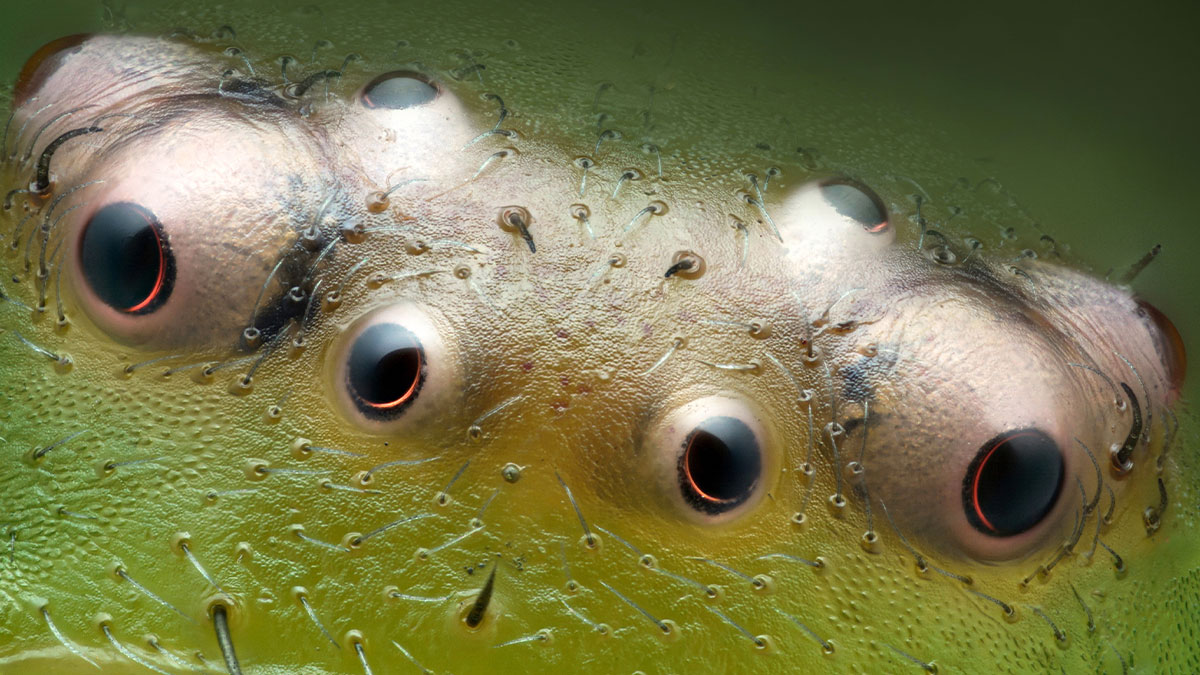
Bedlno, Świętokrzyskie, Poland
"Eyes of green crab spider (Diaea dorsata)."
That looks like something from the more spooky version of Monsters, Inc.
Verona, Veneto, Italy
"Electrical arc between a pin and a wire."
looks like a ufo sending a beam down while electrocuting the ground
Columbia University
Department of Neurobiology and Behavior
New York, New York, USA
"Cluster of octopus (Octopus hummelincki) eggs."
Vernal, Utah, USA
"Agatized dinosaur bone."
Mannheim, Baden-Wuerttemberg, Germany
"Golden bug eggs on a sage leaf."
I especially like the surface of the sage leaf. You can feel it is velvety to the touch, and this picture shows exactly why that is.
Glendale, Arizona, USA
"Ocelli between the compound eyes of a yellow jacket."
Tübingen, Baden-Württemberg, Germany
"Ink dot on Japanese washi paper."
Helsinki University
Helsinki, Uudenmaan lääni, Finland
"Slime mold (Cribraria cancellata)."
University of Zurich
Department of Molecular Life Sciences
Zurich, Switzerland
"Developing nervous system in the eye of a 7-day-old chick embryo."
Maria Enzersdorf, Austria
"Cross section of European beach grass (Ammophila arenaria) leaf."
Aywaille, Liege, Belgium
"Vinyl player needle on scratched vinyl disk."
"scratched" as in damaged? I'd like to see how that looks with a pristine vinyl.
Medienbunker Produktion
Bendorf, Rheinland Pfalz, Germany
"Pollen in a garden spider (Araneus) web."
National Institutes of Health
NICHD
Bethesda, Maryland, USA
"Human neurons."
Dallas, North Carolina, USA
"Moss sporophyte with spores (green)."
Dallas NC is just NW of Charlotte, if anyone is wondering. ;)
Oberzent-Airlenbach, Hessen, Germany
"Opening of a hibiscus flower (Hibiscus moscheutos) exposing the pollen in four stages, each ten minutes apart."
Bánd, Veszprém, Hungary
"Slime mold on a rotten twig with water droplets."
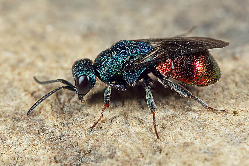
Oberzent-Airlenbach, Hessen, Germany
"Dorsal part of cuckoo wasp (Hedychrum gerstaeckeri) abdomen."
Wloclawek, Kujawko-Pomorskie, Poland
"Water mite (Arrenurus)."
Suwalki, Podlaskie, Poland
"Two water fleas (Daphnia sp.) with embryos (left) and eggs (right)."
Howard Hughes Medical Institute (HHMI), Janelia Research Campus
Ashburn, Virginia, USA
"Aster anther cross-section with pollen grains (green)."
University of Nottingham
School of Life Sciences, Super Resolution Microscopy
Nottingham, Nottinghamshire, United Kingdom
"Dandelion (Traxacum officinale) cross section showing curved stigma with pollen."
Understanding the raw and gritty struggles faced by communities is crucial, much like unveiling intricate details only visible under a microscope. At Kensington Avenue, residents share their poignant stories which reflect societal issues at their core, akin to the way photomicrography opens up hidden worlds of science.
Delve deeper into these revelations about life’s unseen facets by exploring the detailed documentation of raw experiences on the challenges faced by a particular Philadelphia community.
University of Pittsburgh
Department of Ophthalmology
Pittsburgh, Pennsylvania, USA
"Optic nerve head collagen of a pig."
Or, for those of you like me very confused with the title: collagen fibers in the optic nerve head (a structure where the optic nerves exit the eye) in a pig. Collagen is a protein involved in structure and schaffolding. Roughly :)
University of Edinburgh
Institute for Immunology and Infection Research
Edinburgh, MidLothian, United Kingdom
"Acute-stage parasites of Toxoplasma gondii in a human skin cell."
Port Townsend, Washington, USA
"Bract (part of the plant's reproductive structures) of a cannabis plant. The bulbous glands are trichomes."
To simplify: Bracts are modified leaves. The red parts of poinsettias, and the white "petals" of Florida dogwoods are bracts. The flower is actually the little green parts in the middle.
San Anselmo, California, USA
"Seed of a Silene plant."
This is an example of a silene plant Silene-pla...d9-png.jpg 
St. Andrews, Fife, United Kingdom
"Transverse section of rachis (stem) of bracken fern (Pteridium aquilinum)."
Tanta University, Faculty of Science
Department of Zoology
Tanta, Egypt, Arab Republic
"Anterior section of palm weevil."
FP Nature and Landscape Photography
Siracusa, Sicilia, Italy
"Potato tuber sprout."
Is this the lil green bits that pop out when you forget about your potatoes for too long?? 🤔 I didn't know they were so pretty!
University of Oxford
Nuffield Department of Clinical Neurosciences (NDCN)
Oxford, Oxfordshire, United Kingdom
"A network of dopaminergic neurons generated from human stem cells."
MDI Biological Laboratory
Murawala Lab
Bar Harbor, Maine, USA
"Ladybug (Coccinellidae) on a clover (Trifolium repens)."
University of Geneva
Department of Genetics and Evolution
Geneva, Switzerland
"Skin scales of a snake embryo stained with Fast Green dye."
Düsseldorf, Germany
"Cross section of a beach grass (Ammophila arenaria) leaf."
San Anselmo, California, USA
"Slime mold (Lamproderma arcyrioides)."
Lions Eye Institute
Physiology and Pharmacology laboratory
Nedlands, Western Australia, Australia
"Abnormal blood vessel formation in a human retina with severe diabetic retinopathy."
Oregon Department of Agriculture (ODA)
Entomology Lab
Albany, Oregon, USA
"Immature male damselfly (Calopteryx aequabilis)."
Medical College of Georgia at Augusta University
Department of Neuroscience & Regenerative Medicine
Augusta, Georgia, USA
"Differentiated mouse brain tumor cells (actin, microtubules, and nuclei)."
Hounslow, Middlesex, United Kingdom
"Brine shrimp."
Beijing Miteyide Culture Co., Ltd.A925
Beijing, China
"Fiber of nylon stockings."
Johns Hopkins University
Department of Biology
Baltimore, Maryland, USA
"Neuronal axons connecting to the muscles of the iris and the cornea."
Woodend, Waimakiriri, New ZealandA936
"Graffiti from Berlin Wall stone section."
Medical University of South Carolina
Department of Regenerative Medicine & Cell Biology
Charleston, South Carolina, USA
"Section of a small intestine of a mouse."
RBM-CNRS
Montpellier, Herault, France
"Tardigrade (Hypsibius exemplaris)."
Charles University
Department of Experimental Plant Biology
Prague, Czech Republic
"Spores of black truffle (Tuber melanosporum)."
Wow, this looks so 3D! The greens advance and the oranges recede.
Suwalki, Podlaskie, Poland
"Recrystallized mixture of hydroquinone and myoinositol."
Australian National University
Centre for Advanced MicroscopyA306
MacGregor, Australian Capital Territory, Australia
"Autofluorescence in the face of a little two-spotted ladybird (Diomus notescens)."
Club Français de Microscopie
Bailly, France
"Fracture surface of mica (mineral)."
San Anselmo, California, USA
"An insect egg parasitized by a wasp."
Howard Hughes Medical Institute (HHMI), Janelia Research Campus
Ashburn, Virginia, USA
"Floret of a common chicory with pollen grains (spiky balls)."
Woodend, Waimakiriri, New Zealand
"Water mite (Hydrachna sp.)."
Totoro's ghost or Totoro after a CT scan. Totoro-671...7a265e.jpg 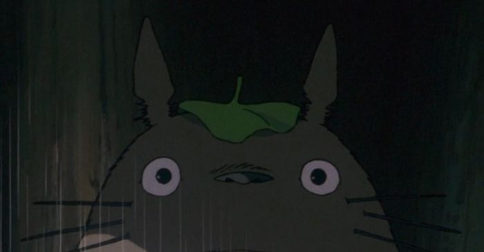
Guangdong Radio and Television
Guangzhou, Guagndong, China
"Stamens of flowers (Anemone cathayensis Kitag. ex Ziman & Kadota)."
Falmouth University
Institute of Photography
Penryn, Cornwall, United Kingdom
"Spores releasing from the sori of a Polypody fern (Polypodium vulgare)."
This be giving off strong "Rat King" energy https://en.wikipedia.org/wiki/Rat_king
Svosov, Zilinsky, Slovak Republic
"Stonewort algae (Chara virgata) reproductive organs - oogonia (female organs) and antheridia (male organs)."
Victoria University of Wellington
School of Biological Sciences; School of Psychology
Wellington, New Zealand
"A neuron densely covered in dendritic spines from the striatum of an adult rat brain."
Saint Lys, Haute-Garonne, France
"Isolated scales on Madagascan sunset moth wing (Chrysiridia ripheus)."
VIB (Flanders Institute of Biotechnology)
Center for Brain and Disease Research
Leuven, Vlaams-Brabant, Belgium
"Fruit fly (Drosophila) brain vasculature."
Amicus Therapeutics
Philadelphia, Pennsylvania, USA
"Dandelion pappus."
University of Turku
Turku Bioscience Centre / Cell Imaging & Cytometry Core and Zebrafish Core
"Gene expression patterns in a drain fly embryo (Clogmia albipunctata) with an open egg. Nutrient storage cells in a tardigrade."
Lovely Professional University
Department of Zoology
Srinagar, Jammu and Kashmir, India
"Small fly killed by 'zombie fly' fungus (Entomophthora muscae)."
Charles University
Department of Experimental Plant Biology
Prague, Czech Republic
"Spores of a black Bagnoli truffle (Tuber mesentericum)."
University of Bordeaux
BioTis-INSERM U1026A383
Pessac, Gironde, France
"Mouse aortic endothelium stained for beta-catenin (green), laminin (purple), smooth muscle actin (red), and Hoechst (cyan)."
University of Helsinki
Individualized Drug Therapy Research Program, Faculty of Medicine, Helsinki, FinlandA700
Helsinki, Finland
"Blood vessels (color gradient) and endothelial cell nuclei (white) in the intestinal villi of a mouse."
CRBM-CNRS
Montpellier, Hérault, France
"Mosquito cells in culture with fluorescent markers for DNA and microtubules."
Hounslow, Middlesex, United Kingdom
"Mosquito larva."
Washington University in St Louis
Department of Medicine - Renal Division, Mahjoub Lab
St Louis, Missouri, USA
"Mouse embryonic kidney showing interstitial fibroblasts (yellow), tubular epithelium (cyan), and nuclei (magenta)."
University of California, San Francisco
Pulmonary, Critical Care, Allergy and Sleep Medicine
San Francisco, California, USA
"Lymphatic vasculature (cyan) and vessels (red) of a mouse lung."
Laval University
Department of Molecular Medicine
Québec, CanadaA858
"Cultured monkey kidney cells labeled for tubulin (blue) and actin (orange) showing pathological accumulation of alpha-syn aggregates (red)."
Medical College of Georgia at Augusta University
Department of Neuroscience & Regenerative MedicineA215
Augusta, Georgia, USA
"Early stage of mouse glioblastoma cell differentiation (actin, microtubules, and mitochondria)."
Max Planck Institute of Molecular Cell Biology and Genetics
Dresden, Saxony, GermanyA903
"Gene expression patterns in a drain fly embryo (Clogmia albipunctata) with an open eggshell."
Oof, I'm trying to get rid of drain flies in my kitchen and bathroom right now. It's not helpful to think of them as pretty!
Yin Works
The Bureau of Microworld Exploration
Beijing, China
"Integrated circuit chip."
Glendale, Arizona, USA
"Mid-tibial tuft on a male orchid bee, used to attract mates."
Oost-Souburg, Zeeland, Netherlands
"Abdominal skin of a tick that engorged with blood."
University of Cape Town
Neuroscience Institute & Department of Human BiologyA249
Cape Town, Western Cape, South Africa
"Astrocytes surrounding a blood vessel in a thin slice of human brain."
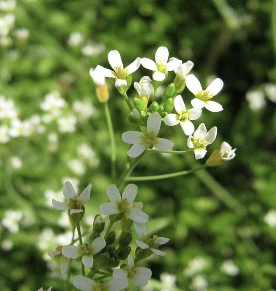
University of Cambridge
Sainsbury Laboratory
Cambridge, Cambridgeshire, United Kingdom
"Leaves arising from thale cress (Arabidopsis thaliana) meristem."
Charité Universitätsmedizin Berlin
Institute of Neurophysiology
Berlin, Germany
"Pyramidal neuron in mouse hippocampus."
Dublin, Ireland
"Larva of a midge fly (Chironomidae)."
Medical University of South Carolina
Department of Regenerative Medicine & Cell Biology
Charleston, South Carolina, USA
"Intestinal villi."
University of Nottingham
School of Life Sciences, Super Resolution MicroscopyA632
Nottingham, Nottinghamshire, United Kingdom
"Malaria parasites and mouse blood cells - tubulin (green), all proteins (purple), DNA (red)."
so interesting ! Thank you so much for this dive into the infinitely small
so interesting ! Thank you so much for this dive into the infinitely small


 No fees, cancel anytime


")
")