167Kviews
Here Are The Best Microscopic Photos, As Announced By The Nikon Small World Photomicrography Competition 2022 (35 Pics)
For the 48th time, Nikon has held its Small World Photomicrography Competition and the winners of the year 2022 have already been announced!
The awards are celebrating the mesmerizing microscopic world and applaud the efforts of those involved with photography, scientists and enthusiasts alike, through the light microscope.
Scroll down for the stunning photographs and don't forget to upvote your favorite ones!
More info: nikonsmallworld.com | Instagram | Facebook | twitter.com
This post may include affiliate links.
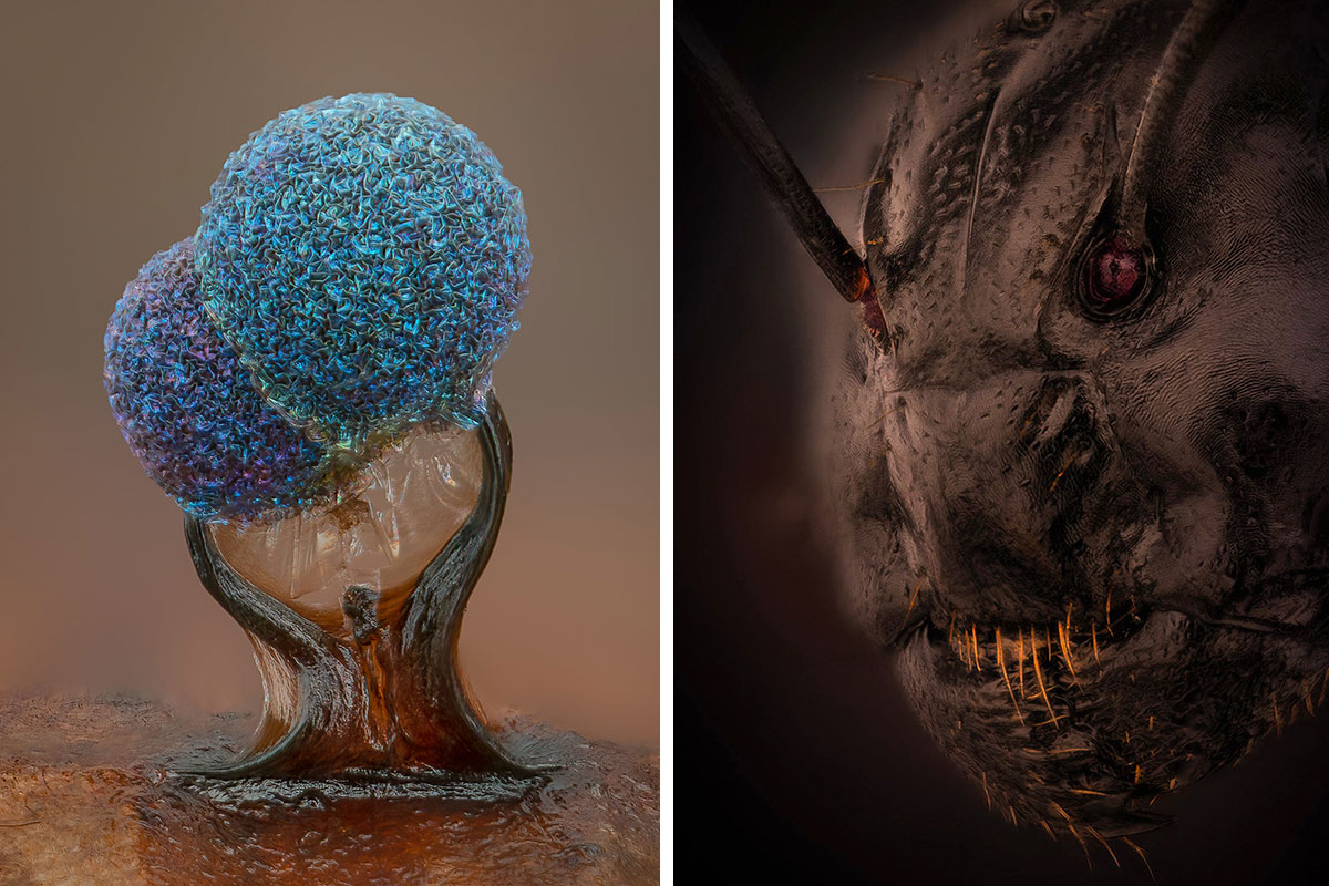
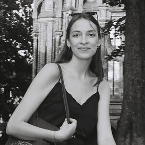
Image Of Distinction - Dr. Eugenijus Kavaliauskas

"Ant (Camponotus)."
Tauragė, Lithuania

The winner of this year's competition is Grigorii Timin, supervised by Dr. Michel Milinkovitch at the University of Geneva with the image of an embryonic hand of a Madagascar giant day gecko. "Masterfully blending imaging technology and artistic creativity, Timin utilized high-resolution microscopy and image-stitching to capture this species of Phelsuma grandis day gecko."
Image Of Distinction - Xinpei Zhang

"Alaskan sand."
Yu Cheng, Ya'an, China
5th Place - Alison Pollack

"Slime mold (Lamproderma)."
San Anselmo, California, USA
According to their website, Nikon’s Small World is regarded as "the leading forum for showcasing the beauty and complexity of life as seen through the light microscope. The Photomicrography Competition is open to anyone with an interest in microscopy and photography."
Image Of Distinction - Yuan Ji

"Butterfly scales."
World Expo Museum
Shanghai, China
Image Of Distinction - Anne-Françoise Tasnier

"Wood cells."
Royal Museum for Central Africa
Department of Wood Biology
Tervuren, Belgium
Besides the photomicrography competition, Nikon is also organizing video competitions, entitled Small World In Motion that "encompass any movie or digital time-lapse photography taken through the microscope."
10th Place - Murat Öztürk

"A fly under the chin of a tiger beetle."
Ankara, Turkey
Honorable Mention - Sebastian Sparenga

"Recrystallized Vitamin C."
McCrone Research Institute
Chicago, Illinois, USA
Let us know what you think of these photographs in the comments!
For more mesmerizing images, check out the winners of previous contests (2016, 2017, 2019, 2020)!
Image Of Distinction - Adolfo Ruiz De Segovia

"Drops of olive oil in water."
Particular
Madrid, Spain
6th Place - Ole Bielfeldt

"Unburned particles of carbon released when the hydrocarbon chain of candle wax breaks down."
Macrofying
Cologne, North Rhine-Westphalia, Germany
Honorable Mention - Ye Fei Zhang

"Butterfly egg."
Jiang Yin, Jiangsu, China
1st Place - Grigorii Timin, Dr. Michel Milinkovitch

"Embryonic hand of a Madagascar giant day gecko (Phelsuma grandis)."
University of Geneva, Geneva, Switzerland
Department of Genetics and Evolution
The Madagascar Giant Day Gecko can grow to be 30ft in length, lives underground, and sends out 3 tentacles from its mouth to grab prey when it senses any kind of vibration coming from above. It can sense anything from pogo sticks to ice machines. Their only natural predators are dynamite and cliffs.
Image Of Distinction - Dr. Stephen S. Nagy

"Diatoms arranged in an exhibition rosette by Klaus D. Kemp."
Montana Diatoms
Helena, Montana, USA
8th Place - Dr. Nathanaël Prunet

"Growing tip of a red algae."
University of North Carolina at Chapel Hill, Chapel Hill, North Carolina, USA
Department of Biology
Image Of Distinction - Dr. Marko Pende

"Transgenic axolotl (CNP:GFP;β3Tubulin:mCherry) showing components of the nervous system. CNP+ Schwann cells (cyan) and axons (magenta)."
MDI Biological Laboratory
Bar Harbor, Maine, USA
Image Of Distinction - Dr. Andrew Posselt

"Bold jumping spider (Phidippus audax)."
University of California, San Francisco (UCSF)
Department of Surgery
Mill Valley, California, USA
Holy f*****g s**t, that's simultaneously the most beautiful and horrifying thing I've ever seen!
11th Place - Ye Fei Zhang

"Moth eggs."
Jiang Yin, Jiangsu, China
Honorable Mention - Dr. Laurent Formery

"Two-month old juvenile sea star (Patiria miniata)."
University of California, Berkeley, Berkeley, California, USA
Department of Molecular and Cell Biology
Image Of Distinction - Karl Deckart

"Dental drill bit studded with diamond chips."
Eckental, Bavaria, Germany
Image Of Distinction - Ahmad Fauzan

"Black and white human hair."
Macro Depok (MD)
Department of Engineering
Jakarta, Indonesia
Image Of Distinction - Yoshihiro Tamaru

"Tail of a planktonic crustacean (Oithona brevicornis)."
Hino, Tokyo, Japan
Image Of Distinction - Gabriel Fernández Fernández Jorge Alberto

"Four o'clock flower (Mirabilis jalapa)."
San Luis, Argentina
Image Of Distinction - Michael Landgrebe

"Moss spore capsule (sporangium)."
Berlin, Germany
Image Of Distinction - Dr. John Hart

"Amino acid crystals (L-glutamine and beta-alanine)."
University of Colorado Boulder
Department of Atmospheric and Oceanic Sciences
Boulder, Colorado, USA
Image Of Distinction - Teresa Zgoda

"Eyeshadow cosmetic."
Arvada, Colorado, USA
Image Of Distinction - Yousef Al Habshi

"Red speckled jewel beetle (Chrysochroa buqueti rugicollis)."
Abu Dhabi, United Arab Emirates
Honorable Mention - Alison Pollack

"Slime mold (Didymium clavus)."
San Anselmo, California, USA
Image Of Distinction - Dr. Keat Ying Chan

"Epithelial cells of a palmskin zebrafish larva."
Academia Sinica
Chen-Hui Chen's Lab
Institute of Cellular and Organismic Biology
Taipei, Taiwan
13th Place - Randy Fullbright

"Agatized dinosaur bone."
Fullbright Studio
Vernal, Utah, USA
16th Place - Dr. Olivier Leroux

"Longitudinal section through a white asparagus shoot tip."
Ghent University, Ghent, Oost-Vlaanderen, Belgium
Department of Biology & Department of Plants and Crops
Honorable Mention - Dr. Igor Siwanowicz

"Radula (rasping tongue) of a marine snail (Turbinidae family)."
Howard Hughes Medical Institute (HHMI), Ashburn, Virginia, USA
Janelia Research Campus
Image Of Distinction - Dr. Honor Glenn

"Human lung cell infected with coronavirus."
Arizona State University
Biodesign Institute
Biodesign Imaging Facility, Center for Immunotherapy, Vaccines, and Virotherapy
Tempe, Arizona, USA
Everyone: Wow! That's amazing! Trump: Drink bleach and it will clear that right up.
Image Of Distinction - Anatoly Mikhaltsov

"Cross section of a leaf of dune grass (Ammophila arenaria)."
Children’s Ecological and Biological Center
Department of Botany
Omsk, Russia
4th Place - Dr. Andrew Posselt

"Long-bodied cellar/daddy long-legs spider (Pholcus phalangioides)."
University of California, San Francisco (UCSF), Mill Valley, California, USA
Department of Surgery
Honorable Mention - Karl Gaff

"Midge larva collected from a freshwater pond."
Dublin, Ireland
14th Place - Nadia Efimova

"Differentiated cultured mouse myoblasts with lysosomes (cyan/green), nuclei (yellow), F-actin (magenta)."
Amicus Therapeutics
Philadelphia, Pennsylvania, USA
I don't understand a word of the description but it looks wonderful 😊
7th Place - Dr. Jianqun Gao, Prof. Glenda Halliday

"Human neurons derived from neural stem cells (NSCs)."
University of Sydney, Sydney, New South Wales, Australia
Central Clinical School
Professor Glenda Halliday's Lab
This is what I imagine the ayahuasca universe to look like right before you get to the part where it's just a void with a giant floating eyeball.
Image Of Distinction - Frank Fox

"Hibiscus flower with pollen."
Trier University of Applied Sciences
Konz, Rheinland-Pfalz, Germany
Honorable Mention - Wim Van Egmond

"Larva of an anemone, found in marine plankton."
Micropolitan Museum
Berkel en Rodenrijs, Zuid-Holland, The Netherlands
Honorable Mention - Dr. Andrea Tedeschi

"Murine sensory-motor cortex following mild traumatic brain injury in a transgenic mouse (expressing Thy1-GFP)."
The Ohio State University, Columbus, Ohio, USA
Wexner Medical Center
Department of Neuroscience
Honorable Mention - Jan Rosenboom

"Diatom (Actinoptychus sp.)."
Rostock, Mecklenburg Vorpommern, Germany
When I was 12, I used to get samples of ditch water and had so much fun looking through the microscope at all the tiny creatures!
20th Place - Hui Lin, Dr. Kim Mcbride

"Human cardiomyocytes (heart cells) derived from induced pluripotent stem cells."
Nationwide Children’s Hospital, Columbus, Ohio, USA
Center for Cardiovascular Research
12th Place - Brett M. Lewis

"Autofluorescence of a single coral polyp (approx. 1 mm)."
Queensland University of Technology, Brisbane, Queensland, Australia
Department of Earth and Atmospheric Science
Image Of Distinction - Pablo Piedra

"Stinger of a small paper wasp (Vespidae Protopolybia)."
La Fortuna de San Carlos, Alajuela, Costa Rica
15th Place - Dr. Ziad El-Zaatari

"Cross sections of normal human colon epithelial crypts."
Houston Methodist Hospital
Houston, Texas, USA
Honorable Mention - Gerd Günther

"Young stem of garden bamboo (Fargesia sp.)."
Düsseldorf, Germany
Image Of Distinction - Dr. Julien Resseguier

"Artery of an Atlantic salmon filled with nucleated red blood cells."
University of Oslo
Department of Biosciences / Immunology
Oslo, Viken, Norway
Image Of Distinction - Dr. Olivier Leroux

"Stem section of hemp (Cannabis sativa)."
Ghent University
Ghent, Oost-Vlaanderen, Belgium


3rd Place - Satu Paavonsalo, Dr. Sinem Karaman

"Blood vessel networks in the intestine of an adult mouse."
University of Helsinki, Helsinki, Finland
Individualized Drug Therapy Research Program, Faculty of Medicine
18th Place - Dr. Julien Resseguier

"Network of macrophages (white blood cells) of an adult zebrafish intestine."
University of Oslo, Oslo, Viken, Norway
Department of Biosciences / Immunology
Image Of Distinction - Enrico Bonino

"Winged ant encased in approximately 20 million-year-old Dominican amber."
Liège, Belgium
Honorable Mention -Dr. Amy Engevik

"Intestinal villi (brush border in magenta)."
Medical University of South Carolina
Department of Regenerative Medicine & Cell Biology
Charleston, South Carolina, USA
Image Of Distinction - Layra G. Cintrón-Rivera

"Zebrafish (Danio rerio) embryo head 72 hours after fertilization."
Brown University
Department of Pathology and Laboratory Medicine
Providence, Rhode Island, USA
Image Of Distinction - Charles B. Krebs

"Licomopha diatoms attached to red alga."
Charles Krebs Photography
Issaquah, Washington, USA
Image Of Distinction - Satu Paavonsalo, Dr. Sinem Karaman

"Blood vessels in the diaphragm of a 9-day-old mouse pup."
University of Helsinki
Individualized Drug Therapy Research Program, Faculty of Medicine
Helsinki, Finland
Image Of Distinction - Dr. David Maitland

"Tip of Pampas grass (Cortaderia selloana). Chlorophyll fluoresces red, lignin blue."
Feltwell, Norfolk, United Kingdom
19th Place - Dr. Tagide Decarvalho

"Bacterial biofilm on a human tongue cell."
University of Maryland, Baltimore County (UMBC), Baltimore, Maryland, USA
Keith R. Porter Imaging Facility
just 1 cell on my tongue has that much hair?? tooth brush now makes sense
9th Place - Dr. Marek Sutkowski

"Liquid crystal mixture (smectic Felix 015)."
Warsaw University of Technology, Warsaw, Poland
Institute of Microelectronics and Optoelectronics


Image Of Distinction - Joe Mckellar

"Human lung cell expressing antiviral Mx1 protein (green), microtubules (cyan) and nuclei (orange)."
The National Center for Scientific Research (CNRS)
Université de Montpellier (UM)
Institut de Recherche en Infectiologie de Montpellier (IRIM)
Montpellier, Hérault, France
Image Of Distinction - Henri Koskinen

"Disco fungus (Lachnum clandestinum) growing on a raspberry (Rubus idaeus)."
Helsinki, Uudenmaan lääni, Finland
Image Of Distinction - Dr. Andrew Moore

"3D rendering of the endoplasmic reticulum in a tissue culture cell."
Howard Hughes Medical Institute (HHMI)
Janelia Research Campus
Ashburn, Virginia, USA
Image Of Distinction - Brian J. Ford

"Embryo of a male rat."
Rothay House
Eastrea, Cambridgeshire, United Kingdom
Something I can basically comprehend! This is really interesting for sure
Image Of Distinction - Dr. Philippe P. Laissue

"Living polyps of a reef-building lobe coral (Porites lobata)."
University of Essex
School of Life Sciences
Colchester, Essex, United Kingdom
Image Of Distinction - Nabodita Sinha, Dr. Ashwani Thakur

"Spokes of amino acid."
Indian Institute of Technology Kanpur
Department of Biological Science and Bioengineering
Kanpur, Uttar Pradesh, India
ok so I'm seeing a tiny face, just 2 black dots for eyes, 2 more for ears, and barely a dot for a mouth. it's hair is done pippi longstocking style. then a really thin neck before arms and a skirt. **Am I reading too much into this 1?
Image Of Distinction - Dr. Jason Hill

"Mouse retina."
Thomas Jefferson University
Sidney Kimmel Cancer Center
Philadelphia, Pennsylvania, USA
Honorable Mention - Bre Hewitt

"Migrating human fibroblast stained for the Golgi (orange), the actin cytoskeleton (magenta), and the nucleus (cyan)."
Drexel University, Philadelphia, Pennsylvania, USA
Department of Biology
Image Of Distinction - Dr. Andrew Moore

"Montage of human cells in different stages of mitosis. Chromosomes (orange) and microtubules (white) are shown."
Howard Hughes Medical Institute (HHMI)
Janelia Research Campus
Ashburn, Virginia, USA
Image Of Distinction - Dr. Andrea Tedeschi

"3D imaging of the vasculature network in an adult mouse spinal cord."
The Ohio State University
Wexner Medical Center
Department of Neuroscience
Columbus, Ohio, USA


Image Of Distinction - Dr. Csaba László Pintér

"European pear rust fungus (Gymnosporangium fuscum), colony of ecidio."
Hungarian University of Agriculture and Life Sciences
Georgikon Faculty
Department of Plant Protection
Keszthely, Zala, Hungary
Fascinating to see this from a microscopic view. My parents had a pear tree which eventually needed felling due to some fungus/"leave rust"... even with the bare eye you'd been able to see the protuberances and discoloration on the leaves, but of course never been able to catch all those details 😲!
Image Of Distinction - Dr. Eric Peterman, Jeff Rasmussen

"Nerve network within the skin of zebrafish (Danio rerio) scales. Different colors depict the different planes and depth of the nerves in individual scales."
University of Washington
Department of Biology
Seattle, Washington, USA
someone's pink force field being attack by someone's yellow powers
Image Of Distinction - Dr. Alejandra Bosco Dr. Monica L. Vetter

"Mouse cornea vasculature (arteries, veins and lymphatics)."
University of Utah
Department of Neurobiology
Salt Lake City, Utah, USA
17th Place - Dr. Daniel Wehner Julia Kolb

"Tail fin of a zebrafish larva with peripheral nerves (green) and extracellular matrix (violet)."
Max Planck Institue for the Science of Light, Erlangen, Bayern, Germany
Department of Biological Optomechanics
How many zebrafish do they photograph for this? This is easily the 4th or 5th one on the list. They are clearly the best subjects of award winning micro photography. Interesting! The zebrafish images have all been stunning!
2nd Place - Caleb Dawson

"Breast tissue showing contractile myoepithelial cells wrapped around milk-producing alveoli."
WEHI, The Walter and Eliza Hall Institute of Medical Research, Melbourne, Victoria, Australia
Department of Immunology
Image Of Distinction - Dr. Zhigang Zheng

"Staff sergeant butterfly eggs (Athyma selenophora)."
Zhuhai Photographers Association
Zhuhai, Guangdong, China
Image Of Distinction - Dr. Arandeep S. Dhanda

"Pathogenic Shigella flexneri bacteria spreading outwards from an infected cell."
Simon Fraser University
Burnaby, British Columbia, Canada
Honorable Mention - Dr. Dylan T. Burnette

"A crawling cell."
Vanderbilt University, Nashville, Tennessee, USA
Department of Cell and Developmental Biology
Image Of Distinction - Kamryn Gerner-Mauro Dr. Jichao Chen

"Lung of a 16.5 day old embryonic mouse with airways labeled by SOX2 (pink) and epithelial progenitors by SOX9 (green)."
The University of Texas MD Anderson Cancer Center
Pulmonary Medicine
Houston, Texas, USA
Image Of Distinction - Yousef Al Habshi

"Section of a damselfly (Odonata Neurobasis chinensis)."
Abu Dhabi, United Arab Emirates


Image Of Distinction - Dr. Tong Zhang

"A double nuclei Bovine Pulmonary Artery Endothelial (BPAE) cell."
Northwestern University
Biological Imaging Facility
Evanston, Illinois, USA
Image Of Distinction - Karie Holtermann, Payal Sarkar

"Digital PCR plate set up with RNA extracted from viruses in wastewater sludge (multiplexed with primer probes of SARS-CoV2, spiked BCoV, and PMMoV)."
City of San Jose Regional Wastewater Lab
San Jose, California, USA
Image Of Distinction - Karl Gaff

"Midge fly larva."
Dublin, Ireland
Honorable Mention - Reuben Philip

"A cell with extra centrosomes beginning to divide."
Mount Sinai Hospital, Toronto, Ontario, Canada
Lunenfeld-Tanenbaum Research Institute
Image Of Distinction - Harikumar R. Suma, Prof. Dr. Pierre Stallforth

"Bacterial colony."
Leibniz Institute for Natural Product Research and Infection Biology (Leibniz-HKI)
Department of Paleobiotechnology
Jena, Thuringia, Germany
Image Of Distinction - Nikky Corthout, Jasper Timmerman

"Clustered iPSC derived neurons."
VIB (Flanders Institute of Biotechnology)
Center for Brain and Disease Research
Leuven, Vlaams-Brabant, Belgium
Image Of Distinction - Dr. Carlo Donato Caiaffa De Carvalho, Dr. Richard Finnell, Dr. Bogdan Wlodarcyk, Dr. Linda Lin

"Embryonic mouse."
Baylor College of Medicine
Center for Precision Environmental Health
Houston, Texas, USA
Honorable Mention - Dr. Zhiguo He

"The actomyosin network at the apical pole of human corneal endothelial cells (revealed by immunofluorescence)."
University Jean Monnet
School of Medicine
Saint-Priest-en-Jarez, Rhône-Alpes, France
Image Of Distinction - Dr. Francisco Lázaro-Diéguez

"Liver cells (hepatocytes)."
Albert Einstein College of Medicine
Bronx, New York, USA
Image Of Distinction - Nadia Efimova

"African green monkey kidney cells (COS-7) with Golgi (blue), lysosomes (green), actin cytoskeleton (magenta) and nuclei (yellow)."
Amicus Therapeutics
Philadelphia, Pennsylvania, USA


Image Of Distinction - Layra G. Cintrón-Rivera

"Developing nervous system of a zebrafish (Danio rerio) six days after fertilization."
Brown University
Department of Pathology and Laboratory Medicine
Providence, Rhode Island, USA
Image Of Distinction - Dr. Guillermo López López

"Peruvian lily (Alstroemeria) stamens."
Alicante, Spain
Image Of Distinction - Danny J. Sanchez

"Etch tube in Brazilian quartz with iron oxide staining."
Mineralien LLC
Valley Village, California, USA
Image Of Distinction - Dr. Aurelia Mapps

"Whole-mounted adult mouse heart."
Johns Hopkins University
Department of Cell, Molecular, Developmental Biology and Biophysical Chemistry
Baltimore, Maryland, USA
Image Of Distinction - Dr. Michelle Giedt

"Nurse cell in a developing fruit fly (Drosophila) follicle."
University of Iowa
Department of Anatomy and Cell Biology
Iowa City, Iowa, USA

This is really cool -- the main reason I started checking (and later downloaded) Bored Panda was to come across things like this I never would have seen otherwise. Even the ones the give me the w*****s are still absolutely gorgeous!!!
Wow... BP's censor bot apparently is nuts. Based on this logic, would Willie Nelson's name also be blanked out?
Load More Replies...This is really cool -- the main reason I started checking (and later downloaded) Bored Panda was to come across things like this I never would have seen otherwise. Even the ones the give me the w*****s are still absolutely gorgeous!!!
Wow... BP's censor bot apparently is nuts. Based on this logic, would Willie Nelson's name also be blanked out?
Load More Replies...