Get Premium
Dark mode theme is available exclusively for premium users. Learn more about the benefits of subscribing.
No fees, cancel anytime.

 Dark Mode Ad-Free Browsing Unlimited Content
 Ad-Free Browsing Unlimited Content Dark Mode
Join 1.2 million Panda readers who get the best art, memes, and fun stories every week!

Do you hear the world's smallest violin playing especially for you? Me neither. But that's probably because it's so minute that it's actually mute...
There's a whole other life lurking beyond what our human eyes can see. And the things we can see aren't always as they appear. If you've ever looked through a microscope, you'll know exactly what we mean. Who knew how much was really hiding on the bristles of a toothbrush? Or that grains of salt could pass for intricate artworks worthy of a place on a gallery wall?
People have been sharing exquisite microscopic images online. They give us a glimpse into a fascinating universe hiding right in not-so-plain sight. Bored Panda has put together a list of the best ones. From a flea giving birth to 6 babies, to microplastics hidden in bread, many of these pictures might change your perspective on the world around you.
Keep scrolling, and don't forget to upvote the ones that blow your mind. We also bring you the story of the world's smallest violin, which is only visible under a microscope. You'll find those details between the images.
This post may include affiliate links.
Stop complaining! The world's tiniest violin has arrived...
The world's smallest violin is officially here. But it's so tiny that it can only be seen under a microscope. It's smaller than a speck of dust and thinner than a human hair. And while it might not be able to respond to your dramatic complaints with a smooth rendition of Vivaldi's Four Seasons, it is paving the way for scientists to do new and exciting things in the near future.
Physicists at Loughborough University used nanotechnology to make the little violin. “Though creating the world’s smallest violin may seem like fun and games, a lot of what we’ve learned in the process has actually laid the groundwork for the research we’re now undertaking,” said Professor Kelly Morrison, Head of Physics at Loughborough University in Britain.
“I’m really excited about the level of control and possibilities we have with the set-up,” added Morisson. “I’m looking forward to seeing what I can achieve – but also what everyone else can do with the system.”
That research that's now possible includes things like improving the efficiency of computers to finding new ways of harvesting energy.
"The violin measures 35 microns long and 13 microns wide, with a micron being one millionth of a metre," reports the BBC. "A human hair typically ranges from 17 to 180 microns in diameter, for comparison."
Well I'll be damned...a camel passing through the eye of a needle.
I have trouble getting thread through the eye. This is mighty impressive. HOW???
Yeah, right? And I have seen this somewhere before. Awesome!
Load More Replies...I've seen this before in a museum near Budapest...his work is amazing!!
Hair turns gray or silver because of the pigment (melanin) that gives hair it's coloring decreases as we age
Mine didn't decrease so much as just run out, like printer ink that's fine then splotchy then gone, all on one page.
Load More Replies...I think that's true for polar bears too. Their fur isn't white, it's clear.
Correct, the lack of pigment doesn't just help with camouflage but it also makes fpr better isolation.
Load More Replies...Yeah I heard about that. This is why those with silvery hair seem to have thin, sparse hair.
To create it, a small chip was coated with two layers of gel-like material called a resist. It was then placed under something called the NanoFrazor, which is a nano-sculpting machine.
"The machine uses thermal scanning probe lithography, a technique where a heated, needle-like tip 'writes' highly precise patterns at the nanoscale," explains the BBC. "This allowed the violin design to be etched on to the chip's surface layer... After it was etched, the underlayer of the resist was dissolved to leave behind a violin-shaped hole."
According to the university, a thin layer of platinum was then deposited into the chip. And a final rinse in acetone removed any remaining material to reveal the finished teeny violin.
That is why a paper cut hurts so much, its all the jagged fibers
Load More Replies...Looks like a big-årse hunk of sea salt sitting atop three braided noodles. Or am I just hungry?
The colors are called "pseudo colours"; they are computer-generated and are a standard technique used with SEM images.
The tiny creation involved big work. And it took the research team several months to refine and test different techniques before announcing that they'd finally made the world's smallest "violin." Now that they've laid the groundwork, it takes around three hours to create a violin using the nanolithography system.
The whole point of making the microscopic "instrument" was to test what the university's cutting-edge nanolithography system is capable of. "Our nanolithography system allows us to design experiments that probe materials in different ways – using light, magnetism, or electricity – and observe their responses," Morrison said.
"Once we understand how materials behave, we can start applying that knowledge to develop new technologies, whether it's improving computing efficiency or finding new ways to harvest energy," she added. "But first, we need to understand the fundamental science and this system enables us to do just that."
They're paired, so they're not in ova or men's emissions.
Load More Replies...Amazing! DNA is the densest form of data storage we know about. 20g of DNA could store all of human knowledge!
So you are.....a woman? Isn't that what 'x' means? How did you get them? Which cell, that is?
They might get the cells from the soft tissue inside the mouth. What you see as 'X' are paired chromosomes (you have 23 pairs of chromosomes in each cell, except s***m/eggs, which have no pairs, and red blood cells, which have no chromosomes at all). What we refer to as X and Y chromosomes don't resemble the shape of those letters.
Load More Replies...Dang. There's a kickin' nightclub in my mouth every time I have coffee or cola.
The team said they created the nanoscale violin as a 'playful reference' to the phrase, “Can you hear the world’s smallest violin playing just for you?”
If you've never heard it, it's something said to mock exaggerated complaints or overly dramatic reactions. And it's often accompanied by a hand gesture mimicking someone playing a tiny violin between their thumb and forefinger.
"The expression is thought to have first appeared on television in the 1970s, popularised by the show M*A*S*H, and has remained part of pop culture thanks to appearances in more recent shows like SpongeBob SquarePants," reads the university's site.
This is why kidney stones are so painful. Because of the sharp shape of uric acid.
There's different kinds of kidney stones and even a tiny one can cause a LOT of pain. I had a large obstructing stone in January that I had to have Lithotripsy for after an ER visit. Staghorn stones are the worst. They have spikes that cause damage when they move. You want surgery before they start moving, you DO NOT want to try and pass them!
Load More Replies...🎵 They stab it with their steely knives, but they just can't kíll the beast.
Turn up the Eagles; the neighbors are listening.
Load More Replies...Fleas are uncool if they're anywhere near me. Water- or otherwise.
Load More Replies...They carry the eggs inside of them, in a pouch known as the brood chamber.
Load More Replies...AI Overview: Water fleas, specifically Daphnia, typically carry their eggs in a brood pouch for about 1-4 days, with an average of 3 days.
I would have smash that thing. Anybody getting fleas in their home, knows how hard it is to get rid of them.
That will be five dollars, please. DUbV_fSW4A...699b42.jpg 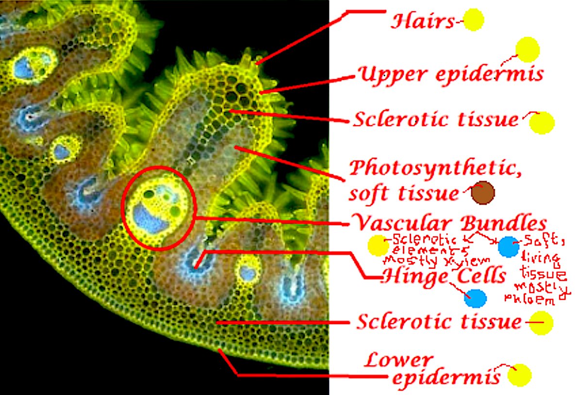
Fun fact: the density of the universe is the same as that of a standard A4 page (80 g/m2).
That's not even close to being true. "Density" of the paper is measured in m² (or cm²), because it's the basic weight/grammage. Unlike the density of the universe, that's measured in cm³ (or m³), because it's not a flat object. For a standard paper with basic weight of 80 g/m² and single A4 sheet being 5 grams, we could calculate it's density to 800 kg/m³ (and that's by a necessary simplification of the whole thing). The density of the universe is about 5,9 protons per m³, which equals 0,0000000000000000000000000099 kg/m³. That's 10^-27 or decimal moved 27 times. Correct me if I'm wrong, because I calculated the paper myself, simplifying it as it would be a cubic object with the same parameters as it has when it's not.
Load More Replies...Of more interest to me is the pixel matrix. Are those half pixels (for better control of intensity) or is it just how they're constructed? Glowing like that, it looks like an OLED to me.
Sunflower (Helianthus annuus, small spiky sphericals, colorized pink), morning glory (Ipomoea purpurea, big sphericals with hexagonal cavities, colorized mint green), hollyhock (Sildalcea malviflora, big spiky sphericals, colorized yellow), lily (Lilium auratum, bean shaped, colorized dark green), primrose (Oenothera fruticosa, tripod shaped, colorized red) and castor bean (Ricinus communis, small smooth sphericals, colorized light green). The image is magnified some x500, so the bean-shaped grain in the bottom left corner is about 50 μm long.
what the actual F**K, that is frozen bubbles under the water not a microscopic image. BP is so s****y now
Have you ever purchased a high-end microscope? I can verify that some of them have a zoom factor so measly that you have to wonder why someone would bother to buy it. Microscopic is a very loose term. Main problem here is that we don't have anything for scale to determine how microscopic the bubble actually are.
Load More Replies...This photo doesn't belong here, as other people have said already. But why the héll are you using it as the thumbnail of this gallery? WTF?
For a second I thought it was baskets of baguettes and buns at the bakery.
0.7mm is 700μm, so the scale line in the picture is at least in the vicinity of correct.
Load More Replies...Are they really plastics? I know there's microplastic everywhere at this point. But those look like any random fibers and are quite visible in those not-so-microscopic, rather very magnified, pictures.
I know bugs are in our veg and fruits. I just choose to not think about it.
Life is life pal. You may not like it but your body is the only home that billions of little creatures will ever know. It’s not gross, it’s a wonder of nature.
Load More Replies...Not much magnification, I can get almost that by careful use of my phone's macro camera with a magnifying glass in front. It would be more interesting to see down into the groove, especially as stereo records wobble the stylus in both dimensions.
In order: acoustic guitar g string: 139x/834x; diatom: 2,085x/5,560x; paramecium: 1,112x/5,560x; human baby tooth: 1,112x/2,085x.
That looks like my archaic engineering electronics lab bread board not too long ago…
I read that as stethoscope and I was all why where you listening to his foot?
Load More Replies...You keep saying that. The answer is no, not a magnifying glass. Microscopes come in various types and in various magnifications. If you agree so obsessed with magnifying glasses I suggest you buy yourself one.
Load More Replies...Too many of these in my yard! The mosquito infestation is terrible this year in NE US with all the rain we've had. Where is the DDT truck when you need it?
Same in the lower midwest, 30 inches so far this year, nothing but tall grass and mosquitos.
Load More Replies...I always heard that too... but then, how do homes, that haven't had any humans in it for years have dust that is inches thick?
Load More Replies...If you're seeing tiny flies around your plants, the soil is likely infested. I tried the rubbing alcohol method to k**l one badly infested plant but to no avail. The larva ate the roots and the dying plant head was just sitting on the soil unattached to anything. It's not really normal and doesn't signify healthy plant life. I have 4 newer plants now since last year. No flies.
My SO has a beautiful garden and is good with plants, but even he has to deal with pests, especially aphids.
Load More Replies...Woah. When you scroll, the image turns monochrome. That's really cool!!!
It's your brain cancelling out all the colours to get a grey. Put the image in the centre and gently shake your phone up and down but keep your eyes fixed, it'll do the same thing.
Load More Replies...You keep saying that. The answer is no, not a magnifying glass. Microscopes come in various types and in various magnifications. If you agree so obsessed with magnifying glasses I suggest you buy yourself one.
Load More Replies...It's a sort of vessel in the orange. The original Reddit links to https://chestofbooks.com/health/herbs/Medicinal-Plants/Chapter-V-Conducting-Tissue.html that explains what it is.
You don't want to know, actually. But there it is. I've seen the visible ones at a friends when she made Minute Maid frozen orange juice.
KiZHXlo Monocular Mircoscope 40-1600X Magnification. I opened each fry to see close the center.
Does it matter though? t's still an interesting picture, so I don't see why you need to comment that everytime 😂
Load More Replies...
Would've liked to see the entire list but not enough to pay for it.
My dad worked for NZ DSIR back in the day, I saw a LOT of electron microscope pictures, he was the photographer.
I remember DSIR. Now CRIs. Did you ever get to request images of anything specific?
Load More Replies...This is a tick. Dead on arrival, cut in half longitudinally, we're looking into the insides of it. Zecke-Laen...aa0352.jpg 
Mostly fascinating, although I'm a bit concerned about what they had to do to get the animal ones.
Would've liked to see the entire list but not enough to pay for it.
My dad worked for NZ DSIR back in the day, I saw a LOT of electron microscope pictures, he was the photographer.
I remember DSIR. Now CRIs. Did you ever get to request images of anything specific?
Load More Replies...This is a tick. Dead on arrival, cut in half longitudinally, we're looking into the insides of it. Zecke-Laen...aa0352.jpg
Mostly fascinating, although I'm a bit concerned about what they had to do to get the animal ones.


 No fees, cancel anytime


")
")