85Kviews
30 Jaw-Dropping Science Images From The 2023 Nikon Small World Photomicrography Competition
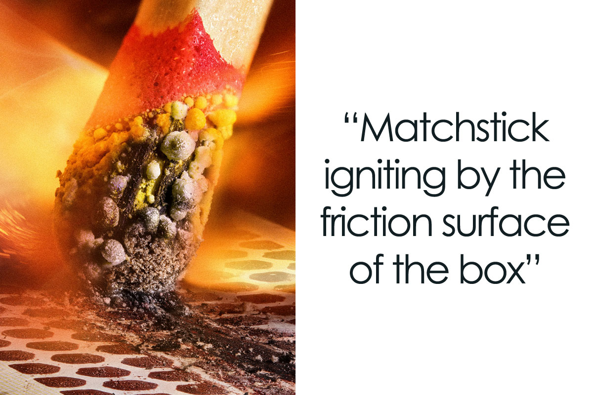
InterviewThe winners of the 49th annual Nikon Small World Photomicrography Competition have been revealed. This year’s first-place prize was awarded to Hassanain Qambari, assisted by Jayden Dickson for his image of a rodent optic nerve head. The second-place winner is Ole Bielfeldt with his image of a matchstick igniting against the surface of a matchbox. Third place was awarded to Malgorzata Lisowska for her image of breast cancer cells.
In addition to the top three winners, the Nikon Small World Photomicrography Competition recognized 83 photos out of nearly 1,900 photo entries from 72 countries.
“The past 49 years of this competition have borne witness to many innovative and pioneering advancements in scientific imaging technology,” Eric Flem, Senior Manager, CRM and Communications at Nikon Instruments, said. “I am consistently awed by how these advancements make it possible to create art out of science for the public to enjoy,” he added.
More info: nikonsmallworld.com | Instagram | twitter.com | Facebook
This post may include affiliate links.
2nd Place - Ole Bielfeldt

Macrofying
Cologne, North Rhine-Westphalia, Germany
"Matchstick igniting by the friction surface of the box."

Nikon Small World is very well known for being the leading forum where people celebrate the skill and beauty of photomicrography.
Competition organizers shared that there are 4 factors to consider when evaluating submissions: originality, informational content, technical proficiency, and visual impact. To select the winners, judges looked at entries that came from all around the globe.
11th Place - Dr. Diego García

Universidad Complutense de Madrid
Real Sociedad Española de Física
Madrid, Spain
"Crystallized sugar syrup."
It looks like a cross between a stack of crumpled paper, and an ocean on a planet with lots of moons.
14th Place - John-Oliver Dum

Medienbunker Produktion
Bendorf, Rheinland Pfalz, Germany
"Sunflower pollen on an acupuncture needle."
The judges of the 2023 Nikon Small World Competition were: Ed Cara, Science and Health Reporter at Gizmodo, James Cutmore, Picture Editor at BBC Science Focus Magazine, Dr. Gary Laevsky, Director of the Confocal Imaging Facility at Princeton University, Dr. Igor Siwanowicz, Research Scientist at Howard Hughes Medical Institute and Dr. Clare Waterman, Cell Biologist and Member of the National Academy of Sciences.
8th Place - Stefan Eberhard

The University of Georgia
Athens, Georgia, USA
"Caffeine crystals."
12th Place - Sherif Abdallah Ahmed

Tanta University
Faculty of Science
Department of Zoology
Tanta, Egypt
"Cuckoo wasp standing on a flower."
I’m absolutely terrified of wasps; the typical “yellow jacket” ones, as it always seems like they want to come after me, LOL. Maybe they smell my high blood glucose? IDK, but I might not be so scared if the local wasps looked as pretty as these little guys! 😂
The Nikon Small World competition was founded in 1974 and in 2011, Nikon Small World in Motion was launched because of advancements in technology that made it possible to record videos or digital time-lapse photos through a microscope.
The 2023 video winners were announced on September 26th.
17th Place - Yuan Ji

World Expo Museum
Shanghai, China
"Chinese moon moth (Actias ningpoana) wing scales."
Here's what they look like when not not magnified moon-moth-...1e1c35.jpg 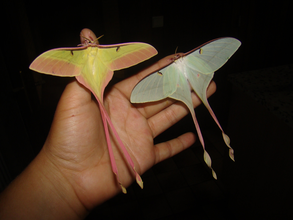
20th Place - Daniel Castranova And Dr. Brant M. Weinstein

National Institutes of Health (NIH)
Eunice Kennedy Shriver National Institute of Child Health and Human Development
Bethesda, Maryland, USA
"Adult transgenic zebrafish head showing blood vessels (blue), lymphatic vessels (yellow), and the skin and scales (magenta)."
Hassanain Qambari, the first-place winner, shared: “The Nikon Small World competition is great, as it showcases amazing work across many disciplines from around the world. All the images presented in the competition represent the beauty and artistic side of science which may otherwise get overlooked. Such a competition not only celebrates the participants' hard work and passion but may also draw and inspire young scientists to pursue a career in STEM. It certainly inspired me.”
Image Of Distinction - Priscilla Vieto Bonilla And Brandon Antonio Segura Torres

Universidad Nacional del Comahue
Department of Biological Sciences
San Carlos de Bariloche, Río Negro, Argentina
"One-week-old Axolotl after hatching."
3rd Place - Malgorzata Lisowska

Independent Value-Based Healthcare Consultant Warsaw, Mazowieckie, Poland
"Breast cancer cells."
Omg. How on earth does something that looks so pretty in an image bring so much pain & sadness?? If no one had said what this was, I’d still be thinking it’s just another cute screensaver pic. Crazy stuff & totally mind-blowing. 🫢
Calling all scientists, photography and video enthusiasts! The Nikon Small World Competition, marking its 50th anniversary in 2024, invites everyone to join in. To be part of this incredible milestone, you can upload your digital images and videos directly on nikonsmallworld.com. Entry forms for Nikon’s 2024 Small World and Small World in Motion Competitions are available at enter.nikonsmallworld.com.
Image Of Distinction - Cagri Yalcin

Impressions Microscopiques
Amsterdam, Noord Holland, The Netherlands
"Crystals of malonic acid dissolved in ethanol."
Image Of Distinction - Walter Machielsen

Rotterdam, Zuid-Holland, The Netherlands
"Buckthorn trichomes."
Image Of Distinction - Danny J. Sanchez

Mineralien LLC
Valley Village, California, USA
"Golden rutile in quartz."
NGL, this looks like it could be a dystopian city in an unknown land! 🤩
Image Of Distinction - Yusuf Ziya Öztürk

TCDD Teknik Müh. Müş. A.Ş.
Ankara, Çankaya, Turkey
"Bee"
4th Place - John-Oliver Dum

Medienbunker Produktion
Bendorf, Rheinland Pfalz, Germany
"Venomous fangs of a small tarantula."
Image Of Distinction - Dr. Frantisek Bednar

Svosov, Zilinsky, Slovakia
"Slime mold (Trichia crateriformis)."
Image Of Distinction - Raghuram Annadana

Raghuram Annadana Photography
Bangalore, Karnataka, India
"Developing stamen and stigma inside a Hibiscus flower bud."
Image Of Distinction - Charles B. Krebs

Charles Krebs Photography
Issaquah, Washington, USA
"Mushroom gills showing sporophores (sporangiophores)."
Image Of Distinction - Dr. Håkan Kvarnström

Bromma, Stockholm, Sweden
"Amoeba (Arcella)."
Honorable Mention - Dr. Amy Engevik

Medical University of South Carolina
Department of Regenerative Medicine & Cell Biology
Charleston, South Carolina, USA
"Neonatal mouse intestinal tissue cells."
Image Of Distinction - Alison Pollack

San Anselmo, California, USA
"Slime mold (Diderma tigrinum)."
Image Of Distinction - Michael Landgrebe

Weissensberg, Bavaria, Germany
"Fossil diatom."
Image Of Distinction - Dr. Pichaya Lertvilai

University of California, San Diego
Scripps Institution of Oceanography
La Jolla, California, USA
"Coral (Acropora granulosa) fluorescing under blue light."
Image Of Distinction - Timothy Boomer

WildMacro
Vacaville, California, USA
"Slime mold (Didymium sp.) fruiting bodies."
1st Place - Hassanain Qambari And Jayden Dickson

Lions Eye Institute
Department of Physiology & Pharmacology
Perth, Western Australia, Australia
"Rodent optic nerve head showing astrocytes (yellow), contractile proteins (red) and retinal vasculature (green)."
Image Of Distinction - Ricardo Roberto Fernández Martínez

IES Virgen de la Luz
Department of Biology and Geology
Avilés, Asturias, Spain
"Tail of planktonic shrimp larvae."
Honorable Mention - Ángel Navarro Gómez

Madrid, Spain
"Carpenter bee (Xylocopa violacea) head and antenna."
But also creepy! Not that bees are, but just the picture!
Load More Replies...Imagine how big raindrops must be when you're that size... especially the really big ones, they must be a real danger
5th Place - Dr. David Maitland

Feltwell, Norfolk, United Kingdom
"Auto-fluorescing defensive hairs covering the leaf surface of Eleagnus angustifolia exposed to UV light."
18th Place - Scott Peterson

New Hope, Minnesota, USA
"A cryptocrystalline micrometeorite resting on a #80 testing sieve."
15th Place - Dr. Pichaya Lertvilai

University of California, San Diego
Scripps Institution of Oceanography
La Jolla, California, USA
"Fluorescent image of an Acropora sp. showing individual polyps with symbiotic zooxanthellae."
13th Place - Satu Paavonsalo And Dr. Sinem Karaman

University of Helsinki
Individualized Drug Therapy Research Program, Faculty of Medicine
Helsinki, Finland
"Blood and lymphatic vasculatures in the ear skin of an adult mouse."
Image Of Distinction - Don Komarechka

Don Komarechka Photography
Ravna Gora, Varna, Bulgaria
"Two fluorescing diamonds."
Image Of Distinction - Foo Yong Ng

Faculty of Science and Technology, Universiti Kebangsaan Malaysia
Department of Biology Science and Biotechnology
Bangi, Selangot, Malaysia
"Moss"
6th Place - Timothy Boomer

WildMacro
Vacaville, California, USA
"Slime mold (Comatricha nigra) showing capillitial fibers through its translucent peridium."
Image Of Distinction - John-Oliver Dum

Medienbunker Produktion
Bendorf, Rheinland Pfalz, Germany
"Cabbage butterfly eggs."
Image Of Distinction - Dr. Saikat Ghosh And Dr. Juan S. Bonifacino

National Institutes of Health (NIH)
NICHD
Bethesda, Maryland, USA
"iPSC-derived human neurons."
Image Of Distinction - Ángel Navarro Gómez

Madrid, Spain
"Mechanosensors in a Venus flytrap."
Image Of Distinction - Frank Reiser

Nassau Community College
Department of Biology
Garden City, New York, USA
"Ostracods and algae (Cladophora)."
Image Of Distinction - Dr. Yu-Hsiu Liu

Academia Sinica
Chen-Hui Chen's Lab in the Institute of Cellular and Organismic Biology
Taipei, Taiwan
"Palmskin zebrafish larva."
Image Of Distinction - Zhao Zengchao

Gaush Meditech Ltd.
Hefei, Anhui Province, China
"Bristle of a millipede (Polyxenidae)."
Image Of Distinction - Ian Gardiner

Calgara, Alberta, Canada
"Clam shrimp (Lynceus mucronatus)."
Image Of Distinction - Dr. Leo Serra

University of Cambridge
Sainsbury Laboratory
Cambridge, Cambridgeshire, United Kingdom
"Bindweed (Convolvulus) leaf epidermal cells autofluorescing."
7th Place - Dr. Grigorii Timin And Dr. Michel Milinkovitch

University of Geneva
Department of Genetics and Evolution
Geneva, Switzerland
"Mouse embryo."
19th Place - Marek Miś

Marek Mis Photography
Suwalki, Podlaskie, Poland
"Stomata in peace lily (Spathiphyllum sp.) leaf epidermis."
Honorable Mention - Dr. Bas Van Bommel

Freie Universität Berlin
Department of Biochemistry
Berlin, Germany
"Rat astrocytes."


Image Of Distinction - Dr. Thomas G.w. Graham

University of California, Berkeley
Department of Molecular and Cell Biology
Berkeley, California, USA
"Algae from a mud puddle."
Image Of Distinction - Taylor Bell

Gustometry + SF Micro Society
Norwalk, Connecticut, USA
"Freshwater amphipod."
Image Of Distinction - Dr. Simon Frederik Merz, Dr. Lea Bornemann And Dr. Ewa Patrycja Smajek

LaVision BioTec, a Miltenyi Biotec Company
Department of Biophysics
Bielefeld, North-Rhine Westphalia, Germany
"Fly (cyan) caught in a Venus flytrap (red)."
Image Of Distinction - Alison Pollack

San Anselmo, California, USA
"Slime mold (Craterium leucocephalum), looking like a beautiful tiny goblet."
Honorable Mention - Dr. Grigorii Timin And Dr. Michel Milinkovitch

University of Geneva
Department of Genetics and Evolution
Geneva, Switzerland
"Dermal collagen in embryonic snake scales."
Honorable Mention - Dr. Arthur Chien And Dr. Ann Na Cho

Macquarie University
Microscopy Facility
Macquarie Park, New South Wales, Australia
"Organ-on-chip system enabling the synaptic conjugation between 3D human embryonic stem cells."
Image Of Distinction - Nikky Corthout And Alex Calzoni

VIB (Flanders Institute of Biotechnology)
Center for Brain and Disease Research
Leuven, Vlaams-Brabant, Belgium
"Retrograde labeled neurons in the cortex of a cortical mouse brain section."
16th Place - Dr. Diego García

Universidad Complutense de Madrid
Real Sociedad Española de Física
Madrid, Spain
"Carbon nanotubes."
Image Of Distinction - Frank Fox

Trier University of Applied Sciences
Konz, Rheinland-Pfalz, Germany
"Marine organism (Pyrocystis lunula, Dinophyceae)."
Image Of Distinction - Sébastien Malo

Saint Lys, Haute-Garonne, France
"Crab spider (Thomisus onustus)."


Image Of Distinction - Sébastien Malo

Saint Lys, Haute-Garonne, France
"Geranium (Geraniaceae) stamen covered in pollen."
Image Of Distinction - Dr. Leo Serra

University of Cambridge
Sainsbury Laboratory
Cambridge, Cambridgeshire, United Kingdom
"Patterns at the surface of an embryonic leaf of Thale cress (Arabidopsis thaliana)."
Honorable Mention - Travis Wagner

Rochester Institute of Technology (RIT)
Department of Mechanical Engineering
Rochester, New York, USA
"Sphagnum moss with two air bubbles on the sample."
Image Of Distinction - Nadia Efimova

Amicus Therapeutics
Philadelphia, Pennsylvania, USA
"Maturing mouse cortical neuron in culture."
Maybe it’s just me, but this pic makes me think of a nighttime view you might see from the view of a plane; showing the powerful lights from whatever large crowded cities are below, even brightly lit roads on the map! With a bit of imagination, I’m sure others could see it, too. 🤩
Image Of Distinction - Charles B. Krebs

Charles Krebs Photography
Issaquah, Washington, USA
"Feeding bryozoan colony zooids. Bryozoans are microscopic aquatic invertebrates that live in colonies."
Image Of Distinction - Dr. Tong Zhang

Northwestern University
Biological Imaging Facility
Evanston, Illinois, USA
"Lily (Lilium) anther cross-section with pollen."
Ultra-slow-motion shot of buckshot beginning to emerge from the barrels of the World's Smallest Double-Barreled Shotgun.
9th Place - Vaibhav Deshmukh

Baylor College of Medicine
Department of Molecular Physiology and Biophysics
Houston, Texas, USA
"Cytoskeleton of a dividing myoblast; tubulin (cyan), F-actin (orange) and nucleus (magenta)."
Image Of Distinction - Dr. Michael John Bridge And Michael Sieverts

University of Utah
HSC Cell Imaging Core
Salt Lake City, Utah, USA
"Mouse femur bone lacunar-canalicular network (voids in bone that house osteocytes and their interconnected micro-tubular processes)."
Image Of Distinction - Dr. David Maitland

Feltwell, Norfolk, United Kingdom
"Wing scales of the cinnabar moth (Tyria jacobaeae) under ultraviolet light (UV)."
Image Of Distinction - Amir Maqbool

Higher Education Department Jammu and Kashmir India
Department of Zoology
Srinagar, Jammu and Kashmir, India
"Phoretic mites on the leg of a bumblebee."


Image Of Distinction - Dr. Andrew M. Posselt

University of California, San Francisco (UCSF)
Division of Transplant Surgery
San Francisco, California, USA
"Blue black weevil (Metapocyrtus sp.)."
That's me before my coffee. Have to say I don't look much better AFTER my coffee . . . .
Honorable Mention - Dr. Andrew M. Posselt

University of California, San Francisco (UCSF)
Division of Transplant Surgery
San Francisco, California, USA
"Underside of cellar spider (Pholcus phalangioides)."

Image Of Distinction - Arturo Calderón, Dr. Miguel Tapia-Rodríguez And Dr. Juan Pedro Laclette

National Autonomous University of Mexico (UNAM)
Department of Immunology
México, Mexico
"Muscle architecture of an evaginating tapeworm (Taenia crassiceps cysticercus)."
Image Of Distinction - Dr. Arthur Chien

Macquarie University
Microscopy Facility
Macquarie Park, New South Wales, Australia
"Cleared mouse embryo."
Image Of Distinction - Marek Miś

Marek Mis Photography
Suwalki, Podlaskie, Poland
"Leaf epidermis stomata (Stromanthe sp.)."
Image Of Distinction - Dr. Colin Rogers And Dr. Erica Weekman

University of Kentucky
Sanders Brown Center on Aging
Lexington, Kentucky, USA
"Mouse retina blood vessels (green) astrocytes (red) and microglia (purple)."
Image Of Distinction - Dr. Alexandre Beber

Institute of Biotechnology CAS
Vestec, Central Bohemia, Czech Republic
"Fluorescent actin filaments (yellow) and fluorescent anillin protein (blue) deposited on a glass coverslip after dewetting."
Honorable Mention - Dr. Nathan P. Myhrvold

Modernist Cuisine
Bellevue, Washington, USA
"Trichinella cyst in pork muscle (Trichinella is a parasitic worm known to cause trichinosis)."
Image Of Distinction - Hsuan Chen

Academia Sinica
Chen-Hui Chen’s Lab in the Institute of Cellular and Organismic Biology
Taipei, Taiwan
"In toto image of the skin and mucous cells in a live zebrafish larva."


Image Of Distinction - Dr. Hema Saranya Ilamathi

Université du Québec à Trois-Rivières (UQTR)
Department of Medical biology
Trois-Rivieres, Quebec, Canada
"Distribution of cellular batteries (mitochondria-yellow) along the transport cables (Tubulin-red, actin-cyan) in human fibroblast."
Image Of Distinction - Dr. Robert Markus

University of Nottingham
School of Life Sciences Imaging (SLIM)
Nottingham, USA
"Actin cytoskeleton of bovine pulmonary epithelial cells."
Image Of Distinction - Dr. Lori O'brien

University of North Carolina at Chapel Hill
Department of Cell Biology and Physiology
Chapel Hill, North Carolina, USA
"Embryonic mouse (Mus musculus) kidney showing the collecting duct (blue) and nephron progenitor (yellow) cells."
Came to say "Oh, that looks like a kidney", and then I read the caption.
Image Of Distinction - Jan Rosenboom

Rostock, Mecklenburg Vorpommern, Germany
"Diatoms (single-celled algae) arranged on the head of a pin."
Image Of Distinction - Dr. Tong Zhang

Northwestern University
Biological Imaging Facility
Evanston, Illinois, USA
"Lily (Lilium) anther cross-section with pollen."
10th Place - Melinda Beccari And Dr. Don W. Cleveland

University of California, San Diego
Department of Cellular and Molecular Medicine
La Jolla, California, USA
"Motor neurons grown in microfluidic device for separation of cell bodies (top) and axons (bottom). Green - microtubules; Red - growth cones (actin)."
Image Of Distinction - Daniel B. Hoffman And Dr. Jacob Sorensen

University of Minnesota
Department of Kinesiology
St. Paul, Minnesota, USA
"Rat skeletal muscle fibers with associated neuromuscular junctions (white)."
Image Of Distinction - Dr. Francisco Lázaro-Diéguez

Albert Einstein College of Medicine
Bronx, New York, USA
"Non-parenchymal liver cells."
Image Of Distinction - Dr. Olivier Leroux

Ghent University
Faculty of Sciences/Faculty of Bioscience Engineering
Ghent, Belgium
"Composition of transverse sections of plant organs."
Image Of Distinction - Hans Schoofs

Uppsala University
Department of Immunology, Genetics and Pathology (IGP)
Uppsala, Sweden
"Blood and lymphatic vessels in a mouse diaphragm."


Image Of Distinction - Dr. Florian Alonso

University of Bordeaux
BioTis-INSERM U1026
Pessac, Gironde, France
"Formation of blood vessels (angiogenesis) in the retina from a Lifeact-EGFP newborn mouse."
Image Of Distinction - Stephen Vidman And Dr. Andrea Tedeschi

The Ohio State University
Department of Neuroscience
Columbus, Ohio, USA
"3D capillary network section of the mammalian brain (dentate gyrus of the hippocampus)."


Am I the only one who read all that and just accepted all those words without REALLY knowing what they said lol
Oh come on. Who amongst us has not popped to the shed to take a quick snap of the symbiotic zooxanthellae on Acropora fluorescing? I can barely move for beakers of ethanol dissolving my malonic acid crystals. /s
Load More Replies...Enjoyed these and would like to see more posts like it. I'm just wondering though, who wakes up in the morning and decides to take a extreme close up of slime mold or a rats optic nerve?


