ADVERTISEMENT
Article created by: Hidrėlėy

The winners of the 51st annual Nikon Small World Photomicrography Competition have been announced, showcasing some of the most stunning and detailed images of the microscopic world. From insects to plant structures, these photographs reveal the beauty and complexity of tiny subjects we often overlook in our daily lives. The competition, run by Nikon Instruments, has been celebrating excellence in microscopy and digital imaging for over five decades, inspiring scientists and artists alike.

This year’s top prize went to China’s Zhang You for his incredible image of a rice weevil perched on a grain of rice, wings fully extended. Using a combination of photography skills, careful lighting, and focus stacking, You captured a moment that blends scientific detail with artistic flair. His work reminds us that even the smallest creatures can be fascinating and beautiful when seen up close.

More info: nikonsmallworld.com | Instagram | Facebook | x.com

#1

3rd Place: John-Oliver Dum, Medienbunker Produktion, Bendorf, Rheinland Pfalz, Germany

Yellow pollen grains captured in stunning microscopic images from the 2025 Nikon Small World Competition

"Pollen in a garden spider web."

© John-Oliver Dum | Nikon Small World Report

RELATED:
    #2

    Honorable Mention: Gregory B. Murray, Pritchard, British Columbia, Canada

    Close-up microscopic image showcasing intricate crystal formations from the 2025 Nikon Small World competition entry.

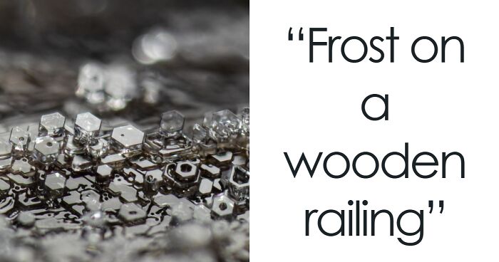
    "Frost on a wooden railing."

    © Gregory B. Murray | Nikon Small World Report

    #3

    Image Of Distinction: Ye Fei Zhang, Jiang Yin, Jiang Su, China

    Close-up microscopic image of two red spiral structures on a textured brown surface from Nikon Small World competition.

    "Butterfly (Artopoetes pryeri) eggs."

    © Ye fei Zhang | Nikon Small World Report

    #4

    Image Of Distinction: Marek Miś, Marek Miś Photography, Suwalki, Podlaskie, Poland

    Microscopic image showcasing colorful crystalline structures with intricate details from the Nikon Small World competition.

    "Crystallized soy sauce."

    © Marek Miś | Nikon Small World Report

    ADVERTISEMENT
    #5

    Image Of Distinction: Dr. David Maitland, Art Of Science, St. Andrews, Fife, United Kingdom

    Microscopic image showing colorful plant cells with distinct blue, green, and red patterns from Nikon Small World competition.

    "Vascular bundles in a bamboo leaf (Phyllostachys sp.)."

    © Dr. David Maitland | Nikon Small World Report

    ADVERTISEMENT
    #6

    2nd Place: Dr. Jan Rosenboom, Rostock, Mecklenburg Vorpommern, Germany

    Microscopic image of a glowing green bubble with intricate patterns captured in the 2025 Nikon Small World competition.

    "Colonial algae (Volvox) spheres in a drop of water."

    © Dr. Jan Rosenboom | Nikon Small World Report

    #7

    5th Place: Dr. Igor Siwanowicz, Howard Hughes Medical Institute (Hhmi), Janelia Research Campus, Ashburn, Virginia, USA

    Colorful microscopic image showcasing intricate details and vibrant structures from the Nikon Small World competition 2025.

    "Spores (blue/purple structures) of a small tropical fern (Ceratopteris richardii)."

    © Dr. Igor Siwanowicz | Nikon Small World Report

    #8

    11th Place: Marek Miś, Marek Miś Photography, Suwalki, Podlaskie, Poland

    Microscopic image showing colorful detailed structures with intricate textures from the 2025 Nikon Small World competition.

    "Sunflower trichomes (hair-like plant outgrowths)."

    © Marek Miś | Nikon Small World Report

    ADVERTISEMENT
    #9

    Honorable Mention: Michael Parra Puentes, Bogotá, Cundinamarca, Colombia

    Close-up microscopic image showing detailed claws with bright orange hairs from the 2025 Nikon Small World Competition.

    "Thoracic and cephalic horn of a male beetle (Golofa porteri)."

    © Michael Parra Puentes | Nikon Small World Report

    ADVERTISEMENT
    #10

    13th Place: Henri Koskinen, Helsinki University, Helsinki, Uudenmaan Lääni, Finland

    Microscopic close-up of red spore capsules with hair-like structures featured in Nikon Small World competition.

    "Slime mold (Arcyria major) releasing spores."

    © Henri Koskinen | Nikon Small World Report

    #11

    14th Place: Manfred Heising, Lwl Museum Of Natural History Münster, Münster, Northrhine-Westphalia, Germany

    Microscopic image of a crystal with intricate yellow fiber-like structures inside, showcasing Nikon Small World competition detail.

    "Quartz with biotic goethite filaments."

    © Manfred Heising | Nikon Small World Report

    #12

    Image Of Distinction: Dr. Gonzalo Quiroga Artigas, Crbm-Cnrs, Montpellier, Herault, France

    Transparent microscopic image of a tardigrade showing internal structures in vibrant red and purple from Nikon Small World competition.

    "Tardigrade."

    © Dr. Gonzalo Quiroga Artigas | Nikon Small World Report

    ADVERTISEMENT
    #13

    Honorable Mention: Michael Robert Peres, Rochester Institute Of Technology, School Of Photographic Arts And Sciences, Rochester, New York, USA

    Microscopic image of a detailed snowflake crystal with intricate patterns from the 2025 Nikon Small World competition.

    "Melting snowflake."

    © Michael Robert Peres | Nikon Small World Report

    ADVERTISEMENT
    #14

    Image Of Distinction: Daniel Evrard, Aywaille, Liege, Belgium

    Close-up microscopic image showing intricate golden and black patterned textures and fine hair-like structures.

    "Androconial (pheromone producing) area of a butterfly (Colias) wing."

    © Daniel Evrard | Nikon Small World Report

    #15

    Image Of Distinction: Dr. Francisco Lázaro-Diéguez, Albert Einstein College Of Medicine, Bronx, New York, USA

    Microscopic image showcasing intricate swirling patterns and vibrant colors from the 2025 Nikon Small World competition.

    "Dedifferentiated liver cell."

    © Dr. Francisco Lázaro-Diéguez | Nikon Small World Report

    #16

    Image Of Distinction: Jianguo Mao, Shanghai, Shanghai, China

    Microscopic image of a colorful water flea with detailed features shown in the 2025 Nikon Small World competition.

    "Pregnant water flea (Daphnia)."

    © Jianguo Mao | Nikon Small World Report

    ADVERTISEMENT
    #17

    1st Place: Zhang You, Kunming, Yunnan, China

    Close-up microscopic image of an insect with wings emerging from a cocoon, showcasing detailed textures and shapes.

    "Rice weevil (Sitophilus oryzae) on a grain of rice."

    © Zhang You | Nikon Small World Report

    ADVERTISEMENT
    #18

    Image Of Distinction: Karl Deckart, Eckental, Bavaria, Germany

    Microscopic image showing vivid rainbow colors forming a sharp triangular pattern from the Nikon Small World competition.

    "Recrystallization of phenyl imidazol."

    © Karl Deckart | Nikon Small World Report

    #19

    Image Of Distinction: Dr. Michael Weber, Berlin Institute Of Health At Charité, Department Of Human Genetics, Berlin, Germany

    Microscopic image revealing intricate organic structure resembling a translucent hand, featured in Nikon Small World competition.

    "Blood vessels in the limb of an embryonic mouse."

    © Dr. Michael Weber | Nikon Small World Report

    #20

    Honorable Mention: Dr. Bruno Cisterna And Dr. Eric Vitriol, Medical College Of Georgia At Augusta University, Department Of Neuroscience & Regenerative Medicine, Augusta, Georgia, USA

    Microscopic image displaying intricate networks of purple and blue neural cells with yellow specks in the Nikon Small World competition.

    "Human neurons reprogrammed from skin cells."

    © Dr. Bruno Cisterna and Dr. Eric Vitriol | Nikon Small World Report

    ADVERTISEMENT
    #21

    Image Of Distinction: Jonathan Muyal, Paris, France

    Colorful abstract microscopic image showcasing intricate geometric patterns from the Nikon Small World competition 2025.

    "Iridescent rutile (mineral) needles in a Burmese ruby."

    © Jonathan Muyal | Nikon Small World Report

    ADVERTISEMENT
    #22

    Image Of Distinction: Marek Miś, Marek Miś Photography, Suwalki, Podlaskie, Poland

    Colorful microscopic image showing bubbles and a heart shape, featured in the 2025 Nikon Small World competition.

    "Air bubbles in melted polyvinyl alcohol."

    © Marek Miś | Nikon Small World Report

    #23

    Image Of Distinction: Dr. Stephen De Lisle, Karlstad University, Department Of Environmental And Life Sciences, Karlstad, Värmland, Sweden

    Microscopic image of intricate patterned seeds with vibrant green and brown textures from the Nikon Small World 2025 competition.

    "Lily flower pollen (autofluorescence)."

    © Dr. Stephen de Lisle | Nikon Small World Report

    #24

    Image Of Distinction: Walter Ferrari, Walter Ferrari Macro, Rio Cuarto, Cordoba, Argentina

    Microscopic close-up of insect eggs with detailed texture and geometric patterns from the Nikon Small World competition.

    "True bug (Hemipteran) eggs on a leaf."

    © Walter Ferrari | Nikon Small World Report

    ADVERTISEMENT
    #25

    16th Place: Rogelio Moreno, Panama, Panama

    Colorful microscopic image showing detailed cellular structures in vibrant green blue orange hues from Nikon Small World competition.

    "Spore sacs (sporangia) of a fern."

    © Rogelio Moreno | Nikon Small World Report

    ADVERTISEMENT
    #26

    Honorable Mention: Jiri Cerny, Institute Of Molecular Genetics Of The Czech Academy Of Sciences, Light Microscopy Core Facility, Prague, Czech Republic

    Close-up microscopic image of a colorful spider highlighting detailed textures from the Nikon Small World competition.

    "Jumping spider."

    © Jiri Cerny | Nikon Small World Report

    #27

    Image Of Distinction: Joe Mckellar, Cnrs, The Institue Of Molecular Genetics Of Montpellier (Igmm), Montpellier, Hérault, France

    Microscopic image showing complex blue cellular structures with intricate textures from Nikon Small World competition 2025.

    "Giant human hepatic cancer cell surrounded by smaller cells."

    © Joe Mckellar | Nikon Small World Report

    #28

    Image Of Distinction: Dr. Mette Handberg-Thorsager, Alexandre Alié And Lisa Maria Ulbrich, Georg-August-University Göttingen, Department Of Multiscale Biology, Göttingen, Niedersachsen, Germany

    Microscopic image showing intricate cellular structures with vibrant red and white details from Nikon Small World competition.

    "Oozoid of a sea squirt (Thalia democratica)."

    © Dr. Mette Handberg-Thorsager, Alexandre Alié and Lisa Maria Ulbrich | Nikon Small World Report

    ADVERTISEMENT
    See Also on Bored Panda
    #29

    Image Of Distinction: Dr. Noah Bressman, Salisbury University, Department Of Biology, Salisbury, Maryland, USA

    Microscopic image of a fish skeleton with vibrant purple and yellow hues from the 2025 Nikon Small World competition.

    "Histologically-stained harvestfish/star butterfish (Peprilus paru)."

    © Dr. Noah Bressman | Nikon Small World Report

    ADVERTISEMENT
    See Also on Bored Panda
    #30

    Image Of Distinction: Solvin Zankl, Kiel, Schleswig-Holstein , Germany

    Close-up of a vibrant, iridescent subject showcasing intricate details in microscopic images from Nikon Small World competition.

    "A floating sea slug (Glaucus atlanticus, also known as the blue sea dragon)."

    © Solvin Zankl | Nikon Small World Report

    #31

    15th Place: Zhang You, Kunming, Yunnan, China

    Microscopic image showing insect wings and abdomen with translucent eggs in the 2025 Nikon Small World competition.

    "Geometer moth (Geometridae) laying eggs."

    © Zhang You | Nikon Small World Report

    ADVERTISEMENT
    See Also on Bored Panda
    #32

    Image Of Distinction: Dr. Laurent Formery, University Of California Berkeley, Department Of Molecular And Cell Biology, Pacific Grove, California, USA

    Microscopic image of a spiky organism with vibrant orange and blue patterns from the 2025 Nikon Small World competition.

    "Skeleton of a juvenile sea cucumber."

    © Dr. Laurent Formery | Nikon Small World Report

    #33

    Image Of Distinction: Stephanie Huang, Victoria University Of Wellington, School Of Biological Sciences; School Of Psychology, Wellington, New Zealand

    Colorful microscopic image showing intricate neurons and their connections in stunning detail from Nikon Small World competition.

    "Pyramidal neurons from the ventral orbital cortex (prefrontal cortex) from an adult rat brain."

    © Stephanie Huang | Nikon Small World Report

    ADVERTISEMENT
    See Also on Bored Panda
    #34

    Image Of Distinction: Bernard Allard, Club Français De Microscopie, Sucy-En-Bry, France

    Microscopic image showing detailed close-up of insect eye and hairs from the 2025 Nikon Small World competition.

    "Parasitic fly (Crataerina hirundinis)."

    © Bernard Allard | Nikon Small World Report

    ADVERTISEMENT
    See Also on Bored Panda
    #35

    Image Of Distinction: Bilal Akhtar, Institute Of Molecular Biology, Neuroscience-Rna Biology, Mainz, Rheinland-Pfalz, Germany

    Microscopic image showing intricate network of colorful neurons and cells with glowing details from Nikon Small World contest.

    "14-day-old mouse neuronal co-culture with astrocytes."

    © Bilal Akhtar | Nikon Small World Report

    #36

    Image Of Distinction: Frederic Labaune, Education Nationale, Auxonne, Burgundy, France

    Close-up microscopic image of vibrant orange fungus resembling a small tree, showcasing intricate details from Nikon Small World competition.

    "Slime mold (Arcyria denudata)."

    © Frederic Labaune | Nikon Small World Report

    #37

    Image Of Distinction: Robert Schmittling, Hillsborough, North Carolina, USA

    Microscopic close-up of a textured surface with a central circular feature from the 2025 Nikon Small World competition.

    "Eye of potato (stomate)."

    © Robert Schmittling | Nikon Small World Report

    ADVERTISEMENT
    ADVERTISEMENT
    See Also on Bored Panda
    #38

    9th Place: Wim Van Egmond, Micropolitan Museum, Berkel En Rodenrijs, Zuid Holland, Netherlands

    Close-up microscopic image showing translucent amber-colored spheres with delicate white filaments in high detail.

    "A fungus (Talaromyces purpureogenus) known for its red, diffused pigment."

    © Wim van Egmond | Nikon Small World Report

    #39

    19th Place: Eduardo Carrasco, Cuenca, Azuay, Ecuador

    Close-up microscopic image of a blue iridescent insect with fungal growth on a green leaf from Nikon Small World competition.

    "Parasitic fungus (Cordycipitaceae) on a fly (Calliphoridae)."

    © Eduardo Carrasco | Nikon Small World Report

    #40

    Honorable Mention: Dr. Frédéric Fercoq And Jean-Gabriel Rothan, Muséum National D'histoire Naturelle, Paris, France

    Microscopic image of a twisting green and blue filament, showcasing details from the Nikon Small World competition.

    "Larvae of a filarial parasite (nematode)."

    © Dr. Frédéric Fercoq and Jean-Gabriel Rothan | Nikon Small World Report

    ADVERTISEMENT
    See Also on Bored Panda
    #41

    Image Of Distinction: Daniel Han, Diatoms Australia (Macro Cosmos Imaging), Sydney, New South Wales, Australia

    Microscopic image of marine diatoms attached to translucent blue structures in vibrant detail from Nikon Small World competition.

    "Diatoms (Arachnoidiscus sp.) on coralline algae."

    © Daniel Han | Nikon Small World Report

    ADVERTISEMENT
    #42

    Image Of Distinction: Doong Yien, Xmato Works, Beijing, China

    Colorful abstract patterns resembling feathers in a microscopic image from the 2025 Nikon Small World competition

    "Crystallization of a mixed solution of alanine and glutamine under polarized light."

    © Doong Yien | Nikon Small World Report

    #43

    Image Of Distinction: Hannah Somers, Mdi Biological Laboratory, Light Microscopy Facility, Bar Harbor, Maine, USA

    Microscopic image of a translucent deep-sea fish illuminated with vibrant pink and blue colors from the Nikon Small World competition.

    "An adult zebrafish showing blood vessels in the brain."

    © Hannah Somers | Nikon Small World Report

    ADVERTISEMENT
    #44

    Image Of Distinction: Heiti Paves, Tallinn, Harju, Estonia

    Microscopic image of a transparent embryo revealing internal organs and structures, featured in Nikon Small World competition.

    "Mouse embryo, sagittal section."

    © Heiti Paves | Nikon Small World Report

    #45

    Image Of Distinction: Dr. Julien Resseguier, University Of Oslo, Department Of Biosciences / Fyscell, Oslo, Viken, Norway

    Close-up microscopic image showcasing intricate cellular structures in vibrant neon colors from the Nikon Small World competition.

    "Immune cells (magenta) protecting the different tissue compartments of the zebrafish intestines."

    © Dr. Julien Resseguier | Nikon Small World Report

    ADVERTISEMENT
    #46

    Image Of Distinction: Shambhavi Dwivedi And Dr. Friedemann Kiefer, University Of Münster, Faculty Of Biology, Muenster, North-Rhine Westphalia, Germany

    Microscopic image showing detailed red and white vascular structures from the 2025 Nikon Small World competition.

    "Mouse lymphatic network (red) flanking blood vessels (white)."

    © Shambhavi Dwivedi and Dr. Friedemann Kiefer | Nikon Small World Report

    #47

    Honorable Mention: Rebecca Lee, Yale University, Department Of Genetrics, New Haven, Connecticut, USA

    Microscopic image highlighting intricate cell structures in vibrant colors from the 2025 Nikon Small World competition.

    "Villi in the mouse small intestine."

    © Rebecca Lee | Nikon Small World Report

    ADVERTISEMENT
    #48

    Image Of Distinction: Dr. Rory L. Cooper And Professor Michel Milinkovitch, University Of Geneva, Department Of Genetics And Evolution, Geneva, Switzerland

    Close-up microscopic image showing glowing orange structures with textured patterns from the 2025 Nikon Small World competition.

    "Wing of the chicken embryo after 11 days of development."

    © Dr. Rory L. Cooper and Professor Michel Milinkovitch | Nikon Small World Report

    #49

    10th Place: Dr. Dylan Burnette And Dr. James Hayes, Vanderbilt University School Of Medicine, Department Of Cell And Developmental Biology, Nashville, Tennessee, USA

    Microscopic image showcasing detailed cell structures with vibrant colors from the 2025 Nikon Small World competition.

    "Heart muscle cells (iPSC-derived) showing condensed chromosomes in metaphase."

    © Dr. Dylan Burnette and Dr. James Hayes | Nikon Small World Report

    ADVERTISEMENT
    #50

    4th Place: Dr. James Hayes, Vanderbilt University, Department Of Cell And Developmental Biology, Nashville, Tennessee, USA

    Microscopic image showing cellular structures with blue fibrous networks and scattered oval-shaped components in Nikon Small World competition.

    "Heart muscle cells with chromosomes condensed following cell division."


    © Dr. James Hayes | Nikon Small World Report

    ADVERTISEMENT