Get Premium
Dark mode theme is available exclusively for premium users. Learn more about the benefits of subscribing.
No fees, cancel anytime.

 Dark Mode Ad-Free Browsing Unlimited Content
 Ad-Free Browsing Unlimited Content Dark Mode
Join 1.2 million Panda readers who get the best art, memes, and fun stories every week!
Do you know what the universal blood type is? Apparently, only one in 10 Americans do. (It’s O-negative, by the way.) How much do you know about the lymphatic system? And do you know if you’re getting enough dietary fiber every day? (Chances are, you aren’t.) The reality is that the human body is a mystery to the majority of us. And even if you’ve been to medical school, there might be plenty of conditions you’ve never encountered.
To learn more about what’s going on in the mysterious world of medicine, we took a trip to Medical Doctors on Instagram. This page shares a variety of photos and information to educate others about rare conditions and provide a glimpse into what's going on inside our hospitals. We’ll warn you right now that some of these images are definitely not for pandas with weak stomachs, and other photos might break your heart. But if you can make it through this list, we hope you’ll learn something new!
This post may include affiliate links.
Little Serafina Murphy pictured only days after serious life-saving heart surgery to fix a hole in her heart at Ann & Robert H. Lurie Children's Hospital of Chicago.
When asked why she was up so soon, her reply is adorable. She replied her Hello Kitty slippers make everything better.
If you're feeling sad, remember this C6 vertebra and how happy it is to hold you (and your head) up every day
Inspiring photo of premature newborn's heartwarming smile ❤️ Without the tube in her nose and the wires on her body, you would have no idea Lauren's daughter was a preemie and spent weeks in the nicu
I think the most beautiful smile that a doctor, a nurse or any health worker can receive is that of a child: it is that smile that answers the question "why I chose this job", is that smile that has not compromised, does not hide anything. It is that smile that is lost between the unconsciousness of not understanding what happens and between the desire to grow, run, jump and play with other children.
The hole that a needle/syringe leaves in the skin as seen by an electron microscope
Please wash your hands!
This is the handprint of an 8 year old boy covered in bacteria.
The boy came in from playing outside, and his mom decided to put his hand print inside a large Petri dish, incubated it for two days, and ended up with a colorful germ garden.
A living ladybug found in the transverse colon during screening colonoscopy
Dirt doesn't stick to scar,
A scar is an area of fibrous tissue that replaces normal skin after an injury. Scars result from the biological process of wound repair in the skin, as well as in other organs and tissues of the body. Because it’s a collagen matrix, it sometimes doesn’t have the same properties as normal skin thus not containing sweat glands and as a result, no dirt/dust sticks to it because the surface of it stays dry.
A gallbladder with multiple gallstones
A patient with severe goiter. A goiter, is a swelling in the neck resulting from an enlarged thyroid gland.
Synapse, the junction between two neurons
I don't think this is a "medical photo" so much as a computer rendering
Pediatric Radiology and Imaging. Designed with kids in mind: Child-friendly Magnetic Resonance Imaging (MRI)
This entry is like a little colourful paradise amidst all the other pictures
Really important to know
Adorable Baby: Jaxon Emmett Buell, defies the odds after being born with only part of a brain and skull. Jaxon Emmett Buell-The Boy Born Without Most Of His Skull Little Jaxon was diagnosed with microhydranencephaly, a type of brain malformation that causes severe intellectual disability and deformed skulls, but in Jaxon’s case, it meant that most of his brain and skull were missing. The condition only affects an estimated one in every 4,859 babies born in the U.S. each year, most of which die shortly after birth. Jaxon’s parents were told of his medical condition before his birth but still decided to carry on the pregnancy. Although Jaxon was not expected to survive very long, he managed to beat all odds and celebrated his first birthday this past September.
Jaxon ended up dying when he was five years old. All types of anencephaly are a tragedy for the whole family. The child generally has no "real" awareness of the world around them and have profound intellectual disabilities (as they are missing all or most of their brain.) They are not "miracles". Even when the children live for years or even decades, they require 24-hr complete care and cannot function or do anything for themselves. If they survive into adulthood, the parents will need to set up a facility or other family member who will be willing to care for the disabled child if/when the parents díe. I do not favor eugenics, but I do support abortion, especially in cases like this. Babies with this severe of a physical disability do not truly get to "live", and put financial and emotional strain on their entire families. Parents need to also consider their existing children (if there are any) and how a profoundly disabled baby who needs 24/7 care would impact the care and attention that the other children in the family receive.
52-year-old woman presented with a painless, gradually enlarging scalp swelling
Filiform warts look different than most warts. They have long, narrow projections that extend from the skin. They can be yellow, brown, pink, or skin-toned. Filiform warts are caused by human papillomavirus (HPV).
Like other warts, the filiform variety is a benign growth that can appear as an individual wart or in a group or cluster. Whereas other warts tend to be either raised or flat growths however, filiform warts tend be quite different in that they are quite long and narrow growths that have a distinctive and undesirable frond like appearance which can be quite distressing, particularly as it favours growing on the face, and more specifically on the lips and eyelids. These warts grow very quickly which is also quite characteristic, as most other warts can take a long time to develop after the initial infection.
Okay, time to tear out my own eyes. I have gotten warts a time or two in my life, always on my toes near my nailbed (so, not plantar warts) and always removed easily with either cryotherapy at the dermatologist or over-the-counter salicylic acid patches. I did not want to know that it was possible to have a wart grow on one's lip.
These blood blister formed due to extreme heat while she was playing basketball for an hour on asphalt on a 100 degree weather day!!!
A blood blister is marked by a raised section of skin filled with blood. They are very similar to blisters caused from friction that fill with a clear fluid. In the case of blood blisters, pressure broke blood vessels and mixed blood with the clear fluid. This combination fills the pocket.
As the feet are filled with many nerves and blood vessels and are under pressure most of the waking day, blisters on the feet can be especially painful.
Depending on where a blister is on the foot, it can be disabling and hard to treat.
Most blisters are harmless and resolve spontaneously, but if they get big, manual drainage or evacuation is the favorable method of treatment.
Open fracture dislocation of the wrist
Oral candidiasis, also known as oral thrush, is a condition in which the fungus Candida albicans accumulates on the lining of the mouth.
Tummy tuck/ Abdminoplasty is not just removing skin/fat!
Sometimes our patients complain more about a belly that still looks "like pregnant" after pregnancies.
This is due to a massive separation of the rectus muscles, a so-called rectus diastasis/ abdominal wall hernia.
This is then repaired by means of two rows of sutures with or without nets (Sublay/ IPOM)
After this „delivery“, the concomitant back pain often disappears because the spine is stabilized by the abdominal wall as well.
This looks horrific and painful. I am not sure I want to go on with this thread.
Anatomy of hand
I need one of those for the shoulder. Mine is messed up and I want to understand it better.
A 12-year-old immigrant female was admitted in the dermatology department with multiple brown lesions on the trunk and face and a large cerebriform plaque on the right side of her scalp.
She was born with multiple brown papules and nodules on the trunk and face and a brown patch on the right side of her scalp. She reported an increase in the size of the lesions on her body and face over time and there was no associated pain or itching. According to history given by her parents, the lesion on the scalp was a small brown flat patch at birth and had increased in size and thickness over the last 8 years and in the past 5 years it had become nodular and cerebriform. The patient complained of intense itching and discharge which had recently become malodorous.
Her birth and developmental history were normal.
In physical examination, her developmental and nutritional status was normal. The head circumference was in normal range and her neurologic, cardiovascular, musculoskeletal, and ophthalmic examinations were normal
The mass on the scalp sized 22 cm × 18 cm × 2.5 cm approximately with a nodular surface spread across the right side of the scalp.
This looks like a case of really, REALLY severe varicose veins.
Why you should never put your feet on a car dashboard.
A woman has sustained horrific injuries after resting her feet on the dashboard of a car. It left her with a crushed hip.
This is what constipation after 19 days looks like. A woman suffering from chronic constipation, discovers her colon severely displaced.
A 15-year-old female presented with a 8 cm x 7 cm, ovoid-shaped, hairy lesion on the right side of her face The palm-sized solitary lesion was present since birth and enlarged as she grew .An interview confirmed no personal or family medical history of melanoma or any form of cancer. The patient denied pain, pruritis, functional problems, or limitations in facial expression due to the lesion
54-year-old man with a history of ulcerative colitis was admitted to the hospital with an intraabdominal abscess. On day 7 of his hospital stay, painful skin lesions developed on his neck
A 57-year-old female presented with a rapidly growing ellipse-shaped nodule on her scalp for two years .The mass did not regress with time and was filled with serous fluid. The patient had a medical history of diabetes and hypertension, and her family history was not significant. Physical examination revealed an oval-shaped mass with a central scar, no discharge, no fluctuation, and painless.
The gross pathological description includes a skin ellipse measuring 1.5 x 1.2 × 0.4 cm with a raised skin lesion and a central crater measuring 1.1 cm.
Microscopically, sections showed skin-abundant pilosebaceous follicles with dilated infundibulum containing lamellated keratin and lined by stratified squamous epithelium with peripheral basal cells and no cytologic atypia
Fascinating to see purified DNA (Deoxyribonucleic acid) inside a test tube
In today’s world of DNA analysis by multiplex and real-time PCR, the importance of high-quality, purified DNA cannot be underestimated. Finding a suitable DNA isolation system to satisfy your downstream application needs is vital for the successful completion of experiments.
There are five basic steps of DNA extraction that are consistent across all the possible DNA purification chemistries: 1) disruption of the cellular structure to create a lysate, 2) separation of the soluble DNA from cell debris and other insoluble material, 3) binding the DNA of interest to a purification matrix, 4) washing proteins and other contaminants away from the matrix and 5) elution of the DNA
Body riddled with parasites as a result of eating raw pork for 10 years.
Trichinellosis, more commonly known as trichinosis, is a parasitic food-borne disease that is caused by eating raw or undercooked meats, particularly pork products infested with the larvae of a type of roundworm called Trichinella.
When a human or animal eats meat that contains infective Trichinella larvae, the acid in the stomach dissolves the hard covering of the cyst around the larvae and releases the worms. The worms pass into the small intestine and, in 1–2 days, become mature. After mating, adult females lay eggs. Eggs develop into immature worms, travel through the arteries, and are transported to muscles. Within the muscles, the worms curl into a ball and encyst (become enclosed in a capsule). The life cycle repeats when meat containing these encysted worms is consumed by another human or animal.
You're not wolves, people. Cook your meat at least a LITTLE. And this is a wolf telling you this, so you know it's legit :p
A 22 year-old male was brought to a clinic for evaluation of intermittent abdominal pain and watery diarrhea of 12 years’ duration. Over the previous 2 months, his symptoms had included vomiting and weight loss. The patient had numerous hyperpigmented macules on his lips, buccal mucosa, fingers, and toes. Computed tomography (CT) and ultrasonography showed duodenojejunal intussusception. Upper gastrointestinal (GI) endoscopy revealed multiple polyps.
Nail clubbing is a deformity of the finger or toe nails associated with a number of diseases, mostly of the heart and lungs.
Nail clubbing occurs when the tips of the fingers enlarge and the nails curve around the fingertips, usually over the course of years. Nail clubbing is sometimes the result of low oxygen in the blood and could be a sign of various types of lung disease.
When it occurs together with joint effusions, joint pains, and abnormal skin and bone growth it is known as hypertrophic osteoarthropathy.
Clubbing is associated with lung cancer, lung infections, interstitial lung disease, cystic fibrosis, or cardiovascular disease.
Ankyloglossia, also known as tongue-tie, is a congenital oral anomaly that may decrease the mobility of the tongue tip and is caused by an unusually short, thick lingual frenulum, a membrane connecting the underside of the tongue to the floor of the mouth. Ankyloglossia varies in degree of severity from mild cases characterized by mucous membrane bands to complete ankyloglossia whereby the tongue is tethered to the floor of the mouth.
My son was tongue tied. He had it "clipped" when he was around 4. The improvement in speech was instant.
An X-ray of conjoined twins born with two backbones and two heads
Conjoined twins are two babies who are born physically connected to each other. Conjoined twins develop when an early embryo only partially separates to form two individuals. Although two babies develop from this embryo, they remain physically connected — most often at the chest, abdomen or pelvis. Conjoined twins may also share one or more internal body organs. Though many conjoined twins are not alive when born (stillborn) or die shortly after birth, advances in surgery and technology have improved survival rates. Some surviving conjoined twins can be surgically separated. The success of surgery depends on where the twins are joined and how many and which organs are shared. It also depends on the experience and skill of the surgical team.
Olecranon bursitis is a condition characterized by swelling, redness, and pain at the tip of the elbow. If the underlying cause is due to an infection, fever may be present. The condition is relatively common and is one of the most frequent types of bursitis.
It usually occurs as a result of trauma or pressure to the elbow, infection, or certain medical conditions such as rheumatoid arthritis or gout.
The underlying mechanism is inflammation of the fluid filled sac between the olecranon and skin.
Chest x-ray of a patient demonstrates a large left sided pulmonary cavity with a small dependent air-fluid level within the left mid zone, in keeping with pulmonary abscess. Patchy airspace opacification more inferiorly within left lower zone.
Scarring/atelectasis within lateral aspect of right upper zone.
A 72-year-old man presented to the emergency department with a 2-day history of an itchy rash on his back. On physical examination, edematous, flagellate plaques and linear patches were present across the patient's entire back and upper buttocks. There was no adenopathy, dermographism, or mucosal involvement
MRI of spinal cord (sagittal view) showing vertebral disc herniation and spinal cord compression at the level of C6-C7
Presented is a case of spinal cord transection, where the spinal cord is cut in half. The spinal cord is the main relay station for both afferent sensory information and efferent motor commands. There are multiple sensory tracts which ascend the spinal cord such as the Dorsal Column-Medial Lemniscus (DCML) and spinothalamic. The DCML carries ipsilateral information of pressure, vibration, fine touch and proprioception from sensory nerve endings in the periphery, whereas the spinothalamic tract carries contralateral information of pain, temperature, pressure and crude touch. The main motor pathway that controls voluntary movement is the lateral corticospinal tract, although there are many others. In cases of spinal cord injury, the spinal cord often goes into spinal shock, which is a characteristic reflexive pattern of areflexia->hyporeflexia->hyperreflexia. The state of a patients reflexes can often be tested clinically by testing a reflex arc such as the patellar reflex. Unfortunately, the patient suffered massive spinal cord trauma, sternal fractures, with bilateral lung contusions leading to a hematothorax and expired
Bobble-head doll syndrome in an infant with an arachnoid cyst
Bobble-head doll syndrome is a rare neurological movement disorder in which patients, usually children around age 3, begin to bob their head and shoulders forward and back, or sometimes side-to-side, involuntarily, in a manner reminiscent of a bobble head doll. The syndrome is related to cystic lesions and swelling of the third ventricle in the brain. Symptoms of bobble-head doll syndrome are diverse but can be grouped into two categories: physical and neurological. The most common form of treatment is surgical implanting of a shunt to relieve the swelling of the brain.
A 1.5-year-old girl presented to the pediatric clinic with the chief complaints of gradual onset excessive head nodding (side-to-side movement) for 3 months. Movements increased with walking, emotions, and stress; decreased during periods of concentration; and were absent during sleep. There were no other complaints or headaches. There was no other significant history.
The child was alert, with normal cognitive function. Neurological examination was normal. Initial laboratory assessment including CBC, hepatic and renal function, and endocrine function tests were normal.
Cranial MRI demonstrated a large left-hemispheric cystic process with a midline shift, well-defined thin-walled suprasellar arachnoid cyst measuring 3 × 5 × 7 cm that obstructed the foramina of Monro, with resulting hydrocephalus ventriculomegaly. Based on the cranial MRI and symptoms, a diagnosis of a suprasellar arachnoid cyst with BHDS was made. The patient underwent endoscopic cystoventriculostomy and cystocisternostomy for the suprasellar arachnoid cyst. During the 6 months of follow-up, the head bobbing disappeared completely, and her growth was normal.
Despite the rareness of bobble-head doll syndrome, it is considered an important condition that must be investigated early to detect the cause and treated promptly to avoid potential complications.
An elevated jugular venous pressure (JVP) is the classic sign of venous hypertension (e.g. right-sided heart failure). JVP elevation can be visualized as jugular venous distension, whereby the JVP is visualized at a level of the neck that is higher than normal. The jugular venous pressure is often used to assess the central venous pressure in the absence of invasive measurements (e.g. with a central venous catheter, which is a tube inserted in the neck veins).
KUB X-ray shows two large and oval radio-opaque shadows in the pelvis
I had a "pinpoint" ischemic stroke over a decade ago that caused permanent visual field loss in my left eye. Immediately following the stroke [not that I knew it was a stroke at the time], I actually lost the "ability" to process colors properly, so I had a weird form of colorblindness (purples and blues looked gray to me, green looked brown/orange, and I couldn't distinguish between shades of red, orange, and yellow at all.) Luckily the color issues resolved months later, but the vision loss remained.
"Lichtenberg Figures", properly known as keraunographic markings, the result of being struck by lightning.
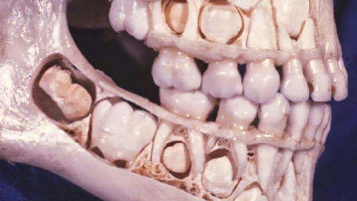
Mulberry molars are a dental condition usually associated with congenital syphilis, characterized by multiple rounded rudimentary enamel cusps on the permanent first molars. Mulberry molars are physically defective permanent molars. The deformity is caused by congenital syphilis. This type of abnormality is characterized by dwarfed molars with cusps covered with globular enamel growths. These teeth are functional but can be cosmetically fixed with crowns, bridges, or implants.
Just above the gum line, the mulberry molar looks normal. A deformity becomes apparent towards the cusp or top grinding surface of the tooth. Here, the size of the mulberry molar is diminished in all aspects, creating a stumpy version of a conventional molar. The cause of the molar atrophy is thought to be enamel hypoplasia, or a deficiency in tooth enamel. The underlying dentin and pulp of the tooth is normal, but the enamel covering or molar sheath is thin and deformed, creating a smaller version of a typical tooth.
The grinding surface of a mulberry molar is also corrupted. Normally, the grinding surface of a molar has a pit and is surrounded by a circular ridge at the top of the tooth, which is used for grinding. The cusp deformity of the mulberry molar is characterized by an extremely shallow or completely absent pit. Instead, the pit area is filled with globular structures bunched together all along the top surface of the cusp. This type of deformity is also thought to be caused by enamel hypoplasia. Mulberry molars are typically functional and do not need treatment. If the deformity is severe or the person is bothered by the teeth, there are several options. The teeth can be covered with a permanent cast crown, stainless steel crown, or the molars can be removed and an implant or bridge can be put in place of the mulberry molar.
Ah yes, this old gem. The "mulberry molars" image where the "molars" are magically located where the incisors usually are XD
Bowel obstruction, also known as intestinal obstruction, is a mechanical or functional obstruction of the intestines which prevents the normal movement of the products of digestion. Either the small bowel or large bowel may be affected. Signs and symptoms include abdominal pain, vomiting, bloating and not passing gas. Mechanical obstruction is the cause of about 5 to 15% of cases of severe abdominal pain of sudden onset requiring admission to hospital.
Causes of bowel obstruction include adhesions, hernias, volvulus, endometriosis, inflammatory bowel disease, appendicitis, tumors, diverticulitis, ischemic bowel, tuberculosis and intussusception. Small bowel obstructions are most often due to adhesions and hernias while large bowel obstructions are most often due to tumors and volvulus. The diagnosis may be made on plain X-rays; however, CT scan is more accurate. Ultrasound or MRI may help in the diagnosis of children or pregnant women.
The condition may be treated conservatively or with surgery. Typically intravenous fluids are given, a tube is placed through the nose into the stomach to decompress the intestines, and pain medications are given. Antibiotics are often given. In small bowel obstruction about 25% require surgery. Complications may include sepsis, bowel ischemia and bowel perforation.
A 27 years male presented with multiple nodular swelling in the bilateral periauricular region with neck
extensions.Scar tissue was present on both the sides with history of previous surgery elsewhere around 17 months back. The swellings were non-tender with mixed consistencies- firm over the nodules but soft in between the nodules. The patient also complained of itching in and around the swelling with visible scratch marks. The patient also gave history of frequent sneezing. watery rhinorrhoea and heaviness of head suggestive of allergic rhinitis
Routine investigation shows significant peripheral eosinophillia and with mild elevation of the ESK.
CT scan of the Paranasal sinuses showed features of bilateral maxillary ethmoidal sinusitis
Hint: Eosinophilic lymphogranuloma
A 44-year-old man with HIV infection presented with a
1-month history of fevers and skin lesions. Examination was notable for blackish-brown lamellated plaques on his limbs and scalp that resembled oyster shells
I posted a comment with info, but BP's new weird AI censorship hid it :/
Lateral view skull radiograph of a patient with multiple myeloma
The classic radiographic appearance of multiple myeloma is that of multiple, small, well-circumscribed, lytic, punched-out, round lesions within the skull, spine, and pelvis. The pattern of lytic or punched-out radiolucent lesions on the skull have been described as resembling raindrops hitting a surface and splashing. How would you manage a patient with multiple myeloma?
A person with a two hour history of phlegmasia cerulea dolens
Phlegmasia cerulea dolens (PCD) is an uncommon severe form of lower extremity deep venous thrombosis (DVT) that obstructs blood outflow from a vein. Upper extremity PCD is less common, occurring in under 10% of all cases. PCD results from extensive thrombotic occlusion of extremity veins, most commonly an "iliofemoral" DVT of the iliac vein and/or common femoral vein. It is a medical emergency requiring immediate evaluation and treatment.
When a thrombus occludes an extremity vein, pressure backs up in the venous system leading plasma fluid to leak out into the interstitium of the affected limb. This increases the pressure of that limb compartment, which can collapse the arteries and lead to acute ischemia, gangrene, hypovolemia, and hemodynamic instability.
Phlegmasia cerulea dolens is best diagnosed with contrast venography, but venous duplex ultrasonography is used more commonly in clinical practice. Magnetic resonance and computed tomography venography can also be used.
The facial nerve is the seventh cranial nerve, or simply CN VII. It emerges from the pons of the brainstem, controls the muscles of facial expression, and functions in the conveyance of taste sensations from the anterior two-thirds of the tongue. The nerves typically travels from the pons through the facial canal in the temporal bone and exits the skull at the stylomastoid foramen.
The facial nerve has five main branches, although the anatomy can vary somewhat between individuals. The branches are, from top to bottom: frontal (or temporal), zygomatic, buccal, marginal mandibular, and cervical. Each of these branches provides input to a group of muscles of facial expression.
Auricular Hematoma or Cauliflower Ear
An auricular hematoma is a collection of blood underneath the perichondrium of the ear and typically occurs secondary to trauma. Auricular deformity, commonly known as "cauliflower ear" is the result of untreated or inadequately treated auricular hematoma.
Fingers necrosis in cold agglutinin disease
Cold agglutinin disease is a rare autoimmune disease characterized by the presence of high concentrations of circulating cold sensitive antibodies, usually IgM and autoantibodies that are also active at body temperatures below 30 °C, directed against red blood cells, causing them to agglutinate and undergo lysis. It is a form of autoimmune hemolytic anemia, specifically one in which antibodies bind red blood cells only at low body temperatures.
According to a post on the American Society of Hematology, this patient received treatment, which helped some of the hemolysis, but unfortunately she did develop gangrene and hand surgeons were contacted to follow-up, which probably means some amputations occurred.
I posted a comment earlier with info, but BP's new weird AI censorship hid it :/
Strawberry tongue seen in scarlet fever.
Scarlet fever is an infectious disease resulting from a group A streptococcus (group A strep) infection, also known as Streptococcus pyogenes. The signs and symptoms include a sore throat, fever, headaches, swollen lymph nodes, and a characteristic rash. The rash is red and feels like sandpaper and the tongue may be red and bumpy. It most commonly affects children between five and 15 years of age.
Scarlet fever affects a small number of people who have strep throat or streptococcal skin infections. The bacteria are usually spread by people coughing or sneezing. It can also be spread when a person touches an object that has the bacteria on it and then touches their mouth or nose. The characteristic rash is due to the erythrogenic toxin, a substance produced by some types of the bacterium. The diagnosis is typically confirmed by culturing the throat.
A patient presented with impaired night vision & a rash on his arm.
"Phrynoderma is a form of follicular hyperkeratosis associated with nutritional deficiency. Phrynoderma is associated with nutritional deficiency, particularly ~ Vitamin A deficiency (commonly), severe malnutrition, Vitamin E and B deficiency, or essential fatty acid deficiency. Phrynoderma is a form of asymptomatic or mildly symptomatic follicular hyperkeratosis, where follicular papules of various sizes with central keratotic plugs that block the follicle openings develop on the skin. New patches may be hypopigmented. Other symptoms of vitamin A deficiency can be present, such as: night blindness and inability to see in bright light."
Tongue necrosis due to Giant cell arteritis (GCA)
Necrosis of the anterior two-thirds of the tongue body. Such lesions are secondary to arteritis of the lingual artery.
Giant cell arteritis (GCA), one of the first causes of arteritis in the elderly, affects the vascular territory of the external carotid artery, especially in its superficial temporal branch1.
A 66-year-old patient was referred for a fever and rapidly progressive deterioration of his general state. The first symptoms appeared several days before, while the patient was visiting Thailand: fever and unusual bilateral headaches were followed by glossodynia and jaw claudication. Within about 2 hours, tongue edema and jaw pain appeared. Intermittent binocular diplopia with transient amaurosis was also noted.
An erythematous and scabbed rash then developed on the patient’s scalp, featuring a necrosis area. The tongue evolved toward cyanosis, further to necrosis. The patient’s case history included bilateral shoulder arthralgia. C-reactive protein was 120 mg/l, and temporal artery biopsy confirmed the diagnosis of GCA. Treatment consisted of high-dose intravenous corticosteroids (methylprednisolone, 1.5 mg/kg/d for 3 days. Articular pain, headaches, and ophthalmologic symptoms drastically improved in < 48 hours, while the scalp lesions responded at a slower rate. Emergency surgery for resection of the tongue necrotic tissue was performed. Because there was satisfactory primary healing, no further treatment was required.
A 50-year-old woman presented to the dental clinic with a 10-day history of tongue and inner cheek pain. She had a history of Crohn’s disease, for which she had been previously treated with prednisone and mesalamine. At this presentation, she had no gastrointestinal or systemic symptoms. On physical examination, she had numerous painful, shallow erosions that merged to form linear “snail track” formations on the dorsal tongue and buccal mucosa. There were no skin lesions. Laboratory studies revealed an absolute eosinophil count of 870 per cubic millimeter (reference range, 50 to 500). A biopsy specimen of the dorsal tongue showed intraepithelial microabscesses with neutrophils and eosinophils, findings consistent with a diagnosis of pyostomatitis vegetans.
Pyostomatitis vegetans is a very rare oral manifestation with unknown pathogenesis. Skin and other mucous membrane involvement may be seen. This lesion has strong association with Inflammatory Bowel Disease (IBD) and may be the first sign of it. The management of Pyostomatitis vegetans is usually based on the management of underlying bowel disease.
The patient was referred for gastroenterologic evaluation, and her bowel disease was found to be quiescent. The oral lesions were treated with topical glucocorticoids and benzocaine. At 1 month of follow-up, her symptoms had resolved, and they did not subsequently recur.
Toxic megacolon is an acute form of colonic distension. It is characterized by a very dilated colon (megacolon), accompanied by abdominal distension (bloating), and sometimes fever, abdominal pain, or shock.
Toxic megacolon is characterized by extreme inflammation and distention of the colon. Common symptoms are pain, distention of the abdomen, fever, rapid heart rate, and dehydration. This is a life-threatening complication that requires immediate medical treatment.
Mastoiditis is the result of an infection that extends to the air cells of the skull behind the ear. Specifically, it is an inflammation of the mucosal lining of the mastoid antrum and mastoid air cell system inside the mastoid process. The mastoid process is the portion of the temporal bone of the skull that is behind the ear. The mastoid process contains open, air-containing spaces. Mastoiditis is usually caused by untreated acute otitis media (middle ear infection).
Cutis verticis gyrata (CVG) is a rare benign cutaneous disorder that is characterized by convoluted folds and deep furrows of the scalp that mimic cerebral sulci and gyri. Cutis verticis gyrata is a medical condition usually associated with thickening of the scalp. The condition is identified by excessive thickening of the soft tissues of the scalp and characterized by ridges and furrows, which give the scalp a cerebriform appearance. Clinically, the ridges are hard and cannot be flattened on applying pressure. Patients show visible folds, ridges or creases on the surface of the top of the scalp. The number of folds can vary from two to roughly ten and are typically soft and spongy. The condition typically affects the central and rear regions of the scalp, but sometimes can involve the entire scalp.
Lamellar ichthyosis, also known as ichthyosis lamellaris and nonbullous congenital ichthyosis, is a rare inherited skin disorder. Affected babies are born in a collodion membrane, a shiny, waxy-appearing outer layer to the skin. This is shed 10–14 days after birth, revealing the main symptom of the disease, extensive scaling of the skin caused by hyperkeratosis. With increasing age, the scaling tends to be concentrated around joints in areas such as the groin, the armpits, the inside of the elbow and the neck. Ichthyosis lamellaris has an autosomal recessive pattern of inheritance.
The picture shows oblong calcific specks in the skeletal muscles parallel to the muscle fibres (rice-grain calcification) consistent with musculoskeletal cysticercosis. The calcifications parallel the long axis of the muscle. This is characteristic of infection with Taenia solium (cysticercosis)
"Taenia solium, commonly known as the pork tapeworm, is a parasitic worm that can cause taeniasis and cysticercosis in humans, leading to significant health issues." Cook your meat thoroughly, everyone!
23-year-old male presented to the emergency department, with rash, mouth sores, and subjective fevers that began after eating fish five days prior. His symptoms started with sores in his mouth and on his lips with penile and anal pruritus. After 24 hours, the patient developed a pruritic rash over his upper extremities, neck, upper back, and palms, as well as two non-painful sores on his penis
On physical examination, he had heme-crusted polycyclic erosions of vermillion lips, buccal mucosa, and labial mucosa.. He was also found to have numerous 2-12 mm erythematous, urticarial, targetoid papules and plaques with central hyperpigmented purple/red duskiness over bilateral palms dorsal hands, upper arms, lateral neck
"Erythema multiforme is an immune-mediated, typically self-limiting, mucocutaneous condition characterised by 'target' lesions. Significant mucosal involvement distinguishes erythema multiforme major from multiforme minor. Episodes can be isolated, recurrent, or persistent. In most cases, erythema multiforme is precipitated by herpes simplex virus (HSV) infection."
Multiple hyperpigmented hyperkeratotic papulonodules over bilateral legs
A 48-year-old man, presented with multiple highly itchy dark raised lesions over bilateral legs.
A long standing case of disseminated hypertrophic Lichen planus treated with oral acitretin
Lichen planus (LP) is a chronic inflammatory and immune-mediated disease that affects the skin, nails, hair, and mucous membranes. It is not an actual lichen, and is only named that because it looks like one. It is characterized by polygonal, flat-topped, violaceous papules and plaques with overlying, reticulated, fine white scale (Wickham's striae), commonly affecting dorsal hands, flexural wrists and forearms, trunk, anterior lower legs and oral mucosa.
Supraclavicular lymph nodes are lymph nodes found above the clavicle, that can be felt in the supraclavicular fossa. The supraclavicular lymph nodes on the left side are called Virchow's nodes. It leads to an appreciable mass that can be recognized clinically, called Troisier sign.
Virchow's node is a lymph node and is a part of the lymphatic system. It is the thoracic duct end node. It receives afferent lymphatic drainage from the left head, neck, chest, abdomen, pelvis, and bilateral lower extremities, which eventually drains into the jugulo-subclavian venous junction via the thoracic duct.
Malignancies of the internal organs can reach an advanced stage before giving symptoms. Stomach cancer, for example, can remain asymptomatic while metastasizing. One of the first visible spots where these tumors metastasize is one of the left supraclavicular lymph node.
Virchow's nodes take their supply from lymph vessels in the abdominal cavity, and are therefore sentinel lymph nodes of cancer in the abdomen, particularly gastric cancer, ovarian cancer, testicular cancer and kidney cancer that has spread through the lymph vessels. Such spread typically results in Troisier's sign, which is the finding of an enlarged, hard Virchow's node.
Cancellous bone, also called spongy bone, is the internal tissue of the skeletal bone. Cancellous bone has a higher surface-area-to-volume ratio than cortical bone and it is less dense. This makes it weaker and more flexible. The greater surface area also makes it suitable for metabolic activities such as the exchange of calcium ions. Cancellous bone is typically found at the ends of long bones, near joints and in the interior of vertebrae. Cancellous bone is highly vascular and often contains red bone marrow where hematopoiesis, the production of blood cells, occurs.
I posted a lot of comments with details and info about the photos listed, since BP once again posted an article with zero context for most of the posts/images. However, due to BP's new weird censor-AI thing, a lot of my info comments were auto-hidden. A few have multiple downvotes now. Try not to downvote them just because they're hidden comments, guys :p Anyway, if you see a comment of mine on one of these images and it's "hidden", it's my research/copy-paste of info related to the image.
Thankyou very much for taking the time to post such interesting comments and explanations
Load More Replies...I posted a lot of comments with details and info about the photos listed, since BP once again posted an article with zero context for most of the posts/images. However, due to BP's new weird censor-AI thing, a lot of my info comments were auto-hidden. A few have multiple downvotes now. Try not to downvote them just because they're hidden comments, guys :p Anyway, if you see a comment of mine on one of these images and it's "hidden", it's my research/copy-paste of info related to the image.
Thankyou very much for taking the time to post such interesting comments and explanations
Load More Replies...


 No fees, cancel anytime


")
")