Weekly The week’s finest content carefully selected to bring you the best articles directly [once per week]
Daily Our top news, meme articles, and a dose of the best content delivered straight to your inbox. [every day]
Funny The funniest articles of the week that had our audience in stitches [once per week]
Animals A delightful mix of top animal photos, memes, and a sprinkle of pet wellness articles [once per week]
Good News A dose of uplifting news delivered directly to your inbox. Pure positivity guaranteed [once per week]
Best Deals Exclusive discounts, our top shopping articles and ever more impressive deals directly to you [once per week]
Art The most captivating photographs, incredible art pieces, and amusing comics—all in one curated collection of the finest art articles [once per week]
Parenting Stories Discover relatable and unbelievable parenting anecdotes from the daily journey of raising a child [once per week]
Brainy Panda The most interesting trivia and quizzes in your inbox [once per week]
BP Daily BP Daily offers readers unbiased, nonpartisan reporting, analysis, and informed opinion in the areas of politics, policy, democracy, ethics, crime, and world news. [once per week]

 Dark Mode


 No fees, cancel anytime

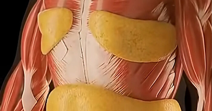
![Body Scans of two Women: 250lb vs 120lb [Pic]](http://lh6.ggpht.com/_9F9_RUESS2E/S0vBbiSEgvI/AAAAAAAACDU/9AzAfMyzlkc/s800/pictureoftheday0007-bodyscans-250-vs-120__700.jpg)
![Body Scans of two Women: 250lb vs 120lb [Pic]](http://lh6.ggpht.com/_9F9_RUESS2E/S0vBbiSEgvI/AAAAAAAACDU/9AzAfMyzlkc/s800/pictureoftheday0007-bodyscans-250-vs-120.jpg "Body Scans of two Women: 250lb vs 120lb [Pic]")


0
2