Get Premium
Dark mode theme is available exclusively for premium users. Learn more about the benefits of subscribing.
No fees, cancel anytime.

 Dark Mode Ad-Free Browsing Unlimited Content
 Ad-Free Browsing Unlimited Content Dark Mode
Join 1.2 million Panda readers who get the best art, memes, and fun stories every week!
20submissions
Finished
Nikon's annual Small World photomicrography competition aims to recognize and showcase the stunning microscopic world that's invisible to the naked eye. Mesmerizing textures, patterns, and colors are revealed in the images taken with the microscopes, offering a peek into a world that we would otherwise miss.
For the 46th time, Nikon has hosted the ever-popular competition, revealing the hidden beauty of the tiny world. Last week, its 20 gorgeous winners were finally unveiled. The top prize this year was snatched by Daniel Castranova, who captured a photo of a juvenile zebrafish with fluorescently "tagged" skeleton, scales, and lymphatic system. The photo was stitched together using more than 350 individual images that were captured with a spinning disc confocal microscope. Scroll down below to see the rest of the winners of the competition, vote for the ones you liked the most, and tell us what you think in the comments down below!
More info: Nikon Small World
Click here & follow us for more lists, facts, and stories.
This post may include affiliate links.
The winning shot of the zebrafish has a significant meaning in the science world, as it helped to make a groundbreaking discovery—zebrafish have lymphatic vessels inside their skull that were previously believed to exist only in mammals. "The image is beautiful, but also shows how powerful the zebrafish can be as a model for the development of lymphatic vessels," said Daniel Castranova, who captured the image. "Until now, we thought this type of lymphatic system only occurred in mammals. By studying them now, the scientific community can expedite a range of research and clinical innovations—everything from drug trials to cancer treatments. This is because fish are so much easier to raise and image than mammals."
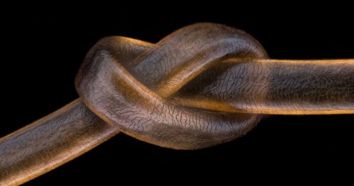
3rd place: Dr. Igor Siwanowicz. Tongue (radula) of a freshwater snail.
9th place: Jason Kirk, Quynh Nguyen. Connections between hippocampal neurons (brain cells).
The winner of the second place is Daniel Knop, who captured and stacked together images of the embryonic development of a clownfish on days 1, 3, 5, and 9. Dr. Igor Siwanowicz, a true veteran of the Small World competition, with a picture of the tongue (radula) of a freshwater snail, was awarded the third place.
13th place: Justin Zoll. Crystals formed after heating an ethanol and water solution containing L-glutamine and beta-alanine.
2nd place: Daniel Knop. Embryonic development of a clownfish (Amphiprion percula) on days 1, 3 (morning and evening), 5, and 9.
20th place: Dr. Dorit Hockman, Dr. Vanessa Chong-Morrison. Skeleton preparation of a short-tailed fruit bat embryo (Carollia perspicillata).
8th place: Dr. Allan Carrillo-Baltodano, David Salamanca. Chameleon embryo (autofluorescence).
7th place: Jason Kirk. Microtubules (orange) inside a cell. Nucleus is shown in cyan.
Talking about the competition, Eric Flem, Communications Manager of Nikon Instruments, said: "For 46 years, the goal of the Nikon Small World competition has been to share microscopic imagery that visually blends art and science for the general public. As imaging techniques and technologies become more advanced, we are proud to showcase imagery that this blend of research, creativity, imaging technology, and expertise can bring to scientific discovery. This year’s first place winner is a stunning example."
4th place: Dr. Vasileios Kokkoris, Dr. Franck Stefani, Dr. Nicolas Corradi. Multi-nucleate spores and hyphae of a soil fungus (arbuscular mycorrhizal fungus).
1st Place: Daniel Castranova, Dr Brant Weinstein & Bakary Samasa. Dorsal view of bones and scales (blue) and lymphatic vessels (orange) in a juvenile zebrafish.
19th place: Dr. Jan Michels. Silica cell wall of the marine diatom Arachnoidiscus sp.
6th place: Dr. Robert Markus, Zsuzsa Markus. Hebe plant anther with pollen.
14th place: Özgür Kerem Bulur. Leaf roller weevil (Byctiscus betulae) lateral view.
15th place: Dr. Eduardo Zattara, Dr. Alexa Bely. Chain of daughter individuals from the asexually reproducing annelid species Chaetogaster diaphanus.
You might also like: 50 ‘Weird Facts’ About The World That Might Give You A Fresh Perspective


 No fees, cancel anytime


")
")


