Get Premium
Dark mode theme is available exclusively for premium users. Learn more about the benefits of subscribing.
No fees, cancel anytime.

 Dark Mode Ad-Free Browsing Unlimited Content
 Ad-Free Browsing Unlimited Content Dark Mode
Join 1.2 million Panda readers who get the best art, memes, and fun stories every week!
33submissions
1week left
Do you know what the universal blood type is? Apparently, only one in 10 Americans do. (It’s O-negative, by the way.) How much do you know about the lymphatic system? And do you know if you’re getting enough dietary fiber every day? (Chances are, you aren’t.) The reality is that the human body is a mystery to the majority of us. And even if you’ve been to medical school, there might be plenty of conditions you’ve never encountered.
To learn more about what’s going on in the mysterious world of medicine, we took a trip to Medical Doctors on Instagram. This page shares a variety of photos and information to educate others about rare conditions and provide a glimpse into what's going on inside our hospitals. We’ll warn you right now that some of these images are definitely not for pandas with weak stomachs, and other photos might break your heart. But if you can make it through this list, we hope you’ll learn something new!
This post may include affiliate links.
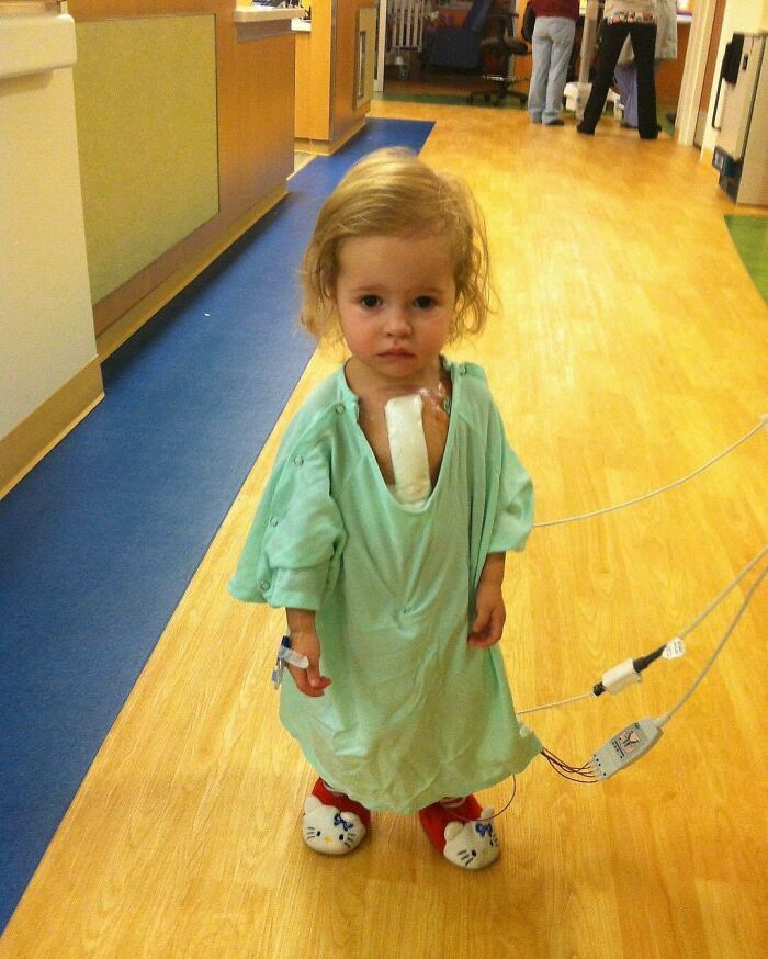
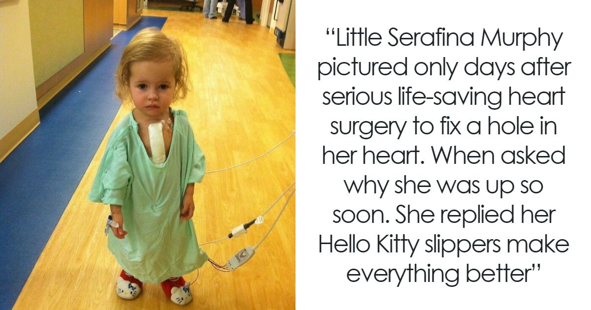
Little Serafina Murphy pictured only days after serious life-saving heart surgery to fix a hole in her heart at Ann & Robert H. Lurie Children's Hospital of Chicago.
When asked why she was up so soon, her reply is adorable. She replied her Hello Kitty slippers make everything better.
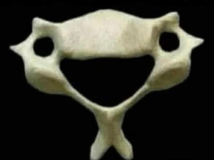
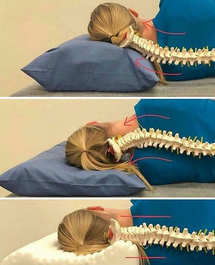
If you're feeling sad, remember this C6 vertebra and how happy it is to hold you (and your head) up every day
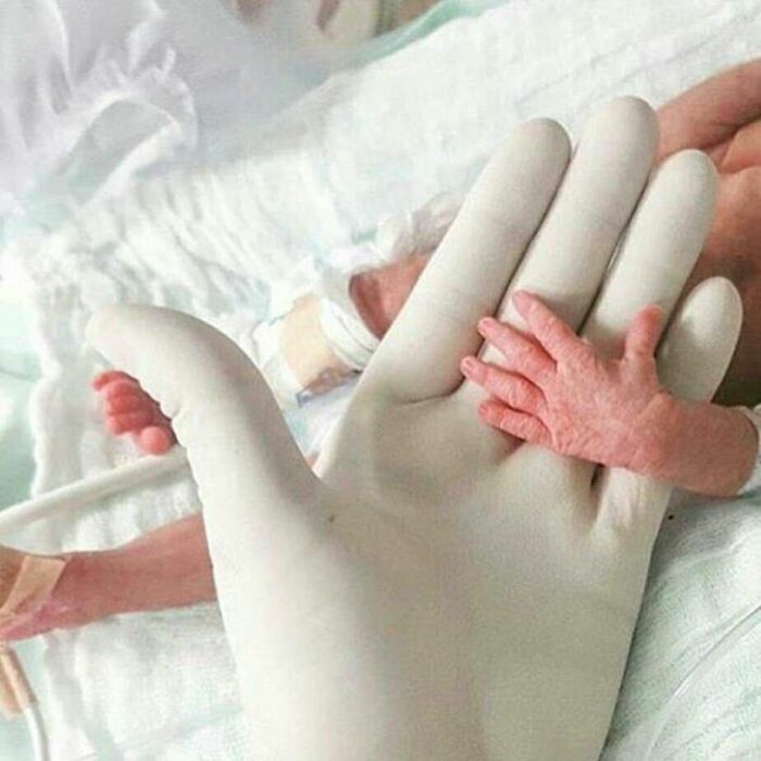
Inspiring photo of premature newborn's heartwarming smile ❤️ Without the tube in her nose and the wires on her body, you would have no idea Lauren's daughter was a preemie and spent weeks in the nicu
I think the most beautiful smile that a doctor, a nurse or any health worker can receive is that of a child: it is that smile that answers the question "why I chose this job", is that smile that has not compromised, does not hide anything. It is that smile that is lost between the unconsciousness of not understanding what happens and between the desire to grow, run, jump and play with other children.
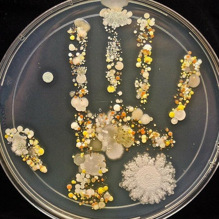
Please wash your hands!
This is the handprint of an 8 year old boy covered in bacteria.
The boy came in from playing outside, and his mom decided to put his hand print inside a large Petri dish, incubated it for two days, and ended up with a colorful germ garden.
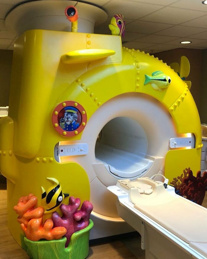
Pediatric Radiology and Imaging. Designed with kids in mind: Child-friendly Magnetic Resonance Imaging (MRI)
A patient with severe goiter. A goiter, is a swelling in the neck resulting from an enlarged thyroid gland.
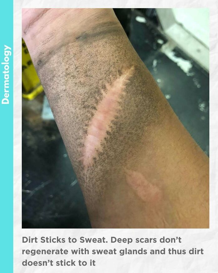
Dirt doesn't stick to scar,
A scar is an area of fibrous tissue that replaces normal skin after an injury. Scars result from the biological process of wound repair in the skin, as well as in other organs and tissues of the body. Because it’s a collagen matrix, it sometimes doesn’t have the same properties as normal skin thus not containing sweat glands and as a result, no dirt/dust sticks to it because the surface of it stays dry.
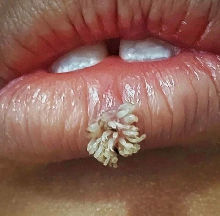
Filiform warts look different than most warts. They have long, narrow projections that extend from the skin. They can be yellow, brown, pink, or skin-toned. Filiform warts are caused by human papillomavirus (HPV).
Like other warts, the filiform variety is a benign growth that can appear as an individual wart or in a group or cluster. Whereas other warts tend to be either raised or flat growths however, filiform warts tend be quite different in that they are quite long and narrow growths that have a distinctive and undesirable frond like appearance which can be quite distressing, particularly as it favours growing on the face, and more specifically on the lips and eyelids. These warts grow very quickly which is also quite characteristic, as most other warts can take a long time to develop after the initial infection.
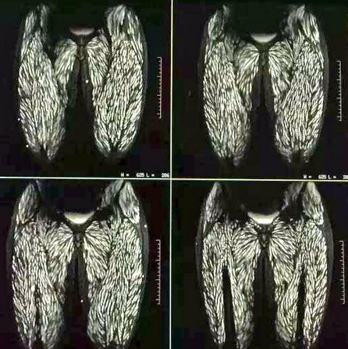
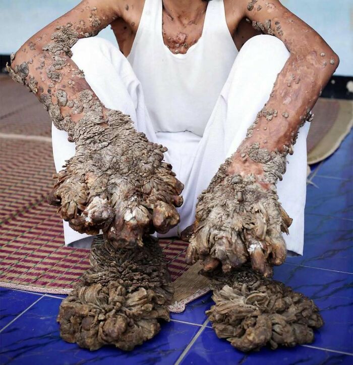
Body riddled with parasites as a result of eating raw pork for 10 years.
Trichinellosis, more commonly known as trichinosis, is a parasitic food-borne disease that is caused by eating raw or undercooked meats, particularly pork products infested with the larvae of a type of roundworm called Trichinella.
When a human or animal eats meat that contains infective Trichinella larvae, the acid in the stomach dissolves the hard covering of the cyst around the larvae and releases the worms. The worms pass into the small intestine and, in 1–2 days, become mature. After mating, adult females lay eggs. Eggs develop into immature worms, travel through the arteries, and are transported to muscles. Within the muscles, the worms curl into a ball and encyst (become enclosed in a capsule). The life cycle repeats when meat containing these encysted worms is consumed by another human or animal.
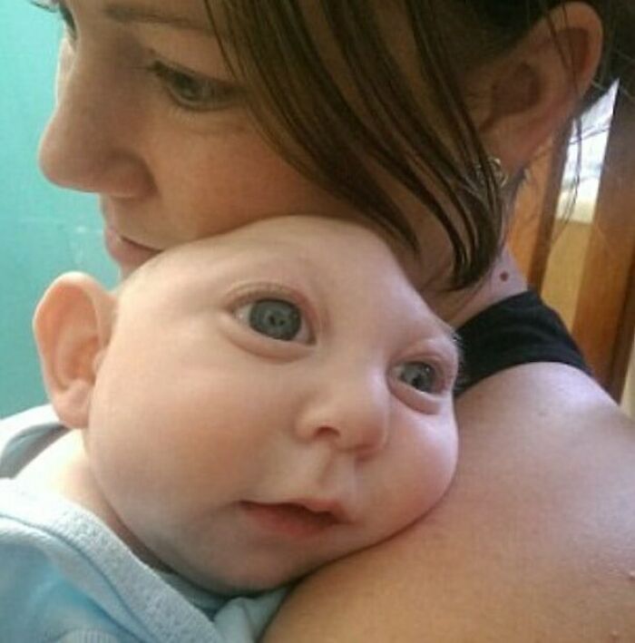
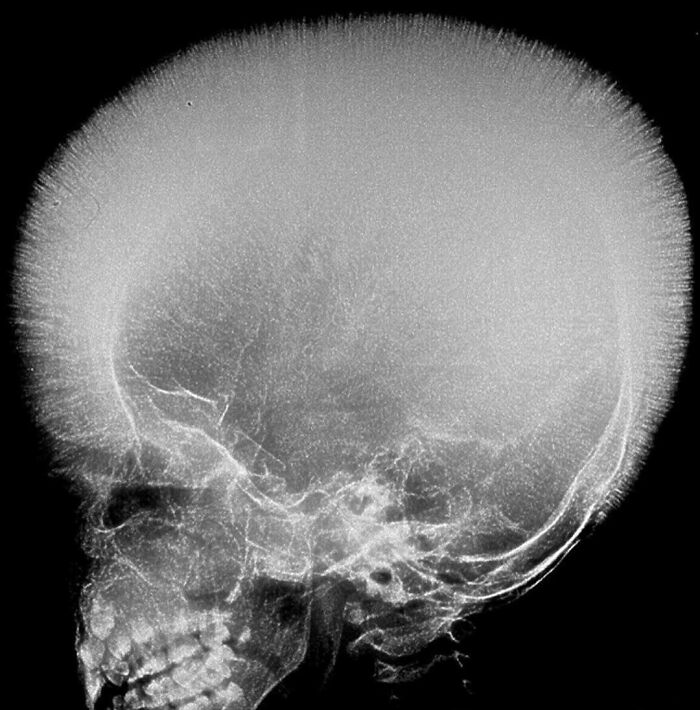
Adorable Baby: Jaxon Emmett Buell, defies the odds after being born with only part of a brain and skull. Jaxon Emmett Buell-The Boy Born Without Most Of His Skull Little Jaxon was diagnosed with microhydranencephaly, a type of brain malformation that causes severe intellectual disability and deformed skulls, but in Jaxon’s case, it meant that most of his brain and skull were missing. The condition only affects an estimated one in every 4,859 babies born in the U.S. each year, most of which die shortly after birth. Jaxon’s parents were told of his medical condition before his birth but still decided to carry on the pregnancy. Although Jaxon was not expected to survive very long, he managed to beat all odds and celebrated his first birthday this past September.
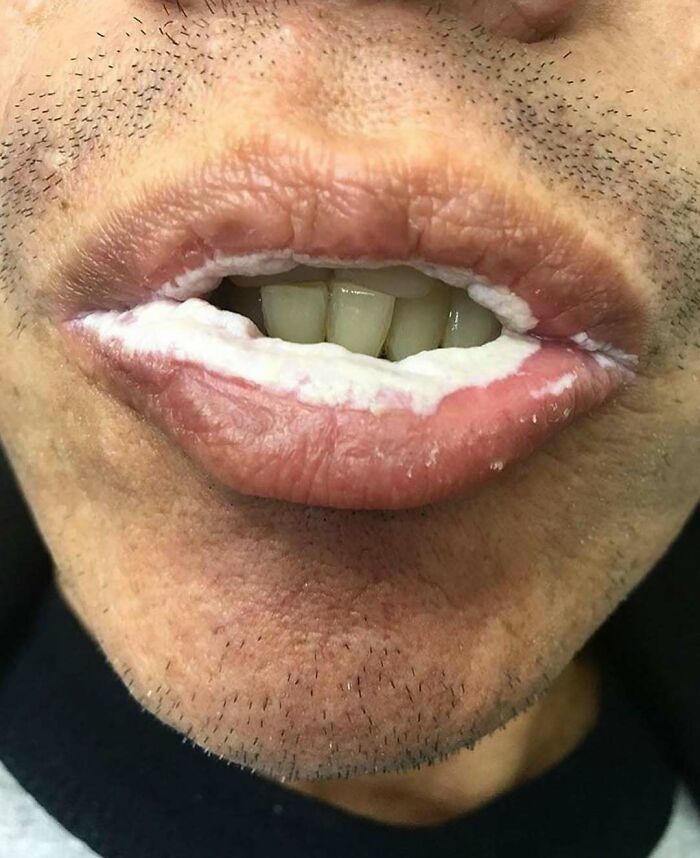
Oral candidiasis, also known as oral thrush, is a condition in which the fungus Candida albicans accumulates on the lining of the mouth.
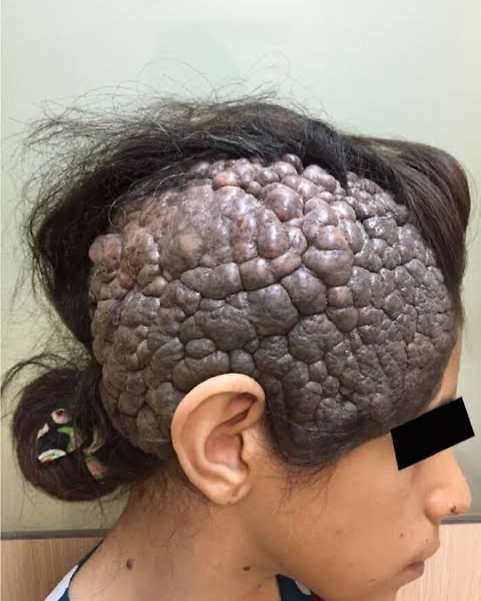
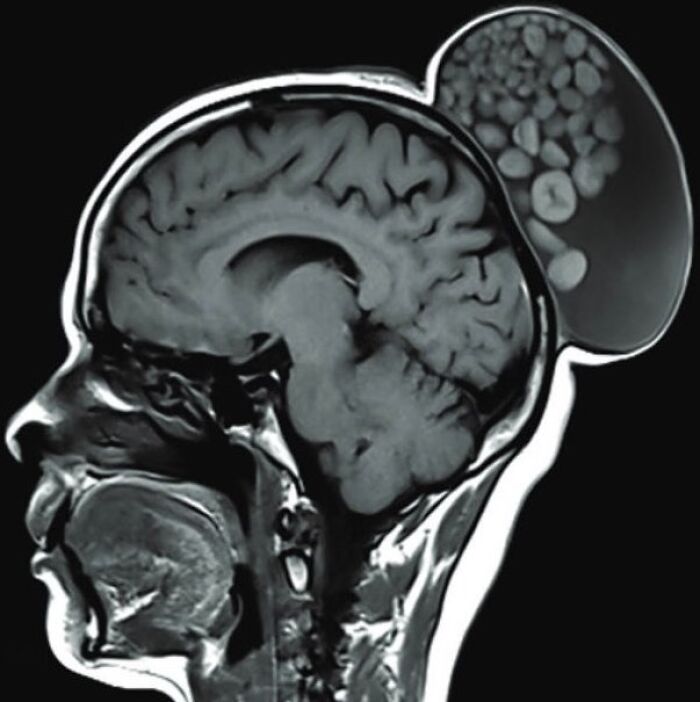
A 12-year-old immigrant female was admitted in the dermatology department with multiple brown lesions on the trunk and face and a large cerebriform plaque on the right side of her scalp.
She was born with multiple brown papules and nodules on the trunk and face and a brown patch on the right side of her scalp. She reported an increase in the size of the lesions on her body and face over time and there was no associated pain or itching. According to history given by her parents, the lesion on the scalp was a small brown flat patch at birth and had increased in size and thickness over the last 8 years and in the past 5 years it had become nodular and cerebriform. The patient complained of intense itching and discharge which had recently become malodorous.
Her birth and developmental history were normal.
In physical examination, her developmental and nutritional status was normal. The head circumference was in normal range and her neurologic, cardiovascular, musculoskeletal, and ophthalmic examinations were normal
The mass on the scalp sized 22 cm × 18 cm × 2.5 cm approximately with a nodular surface spread across the right side of the scalp.
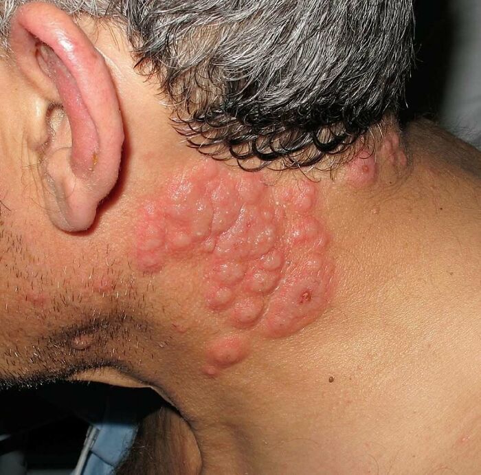
54-year-old man with a history of ulcerative colitis was admitted to the hospital with an intraabdominal abscess. On day 7 of his hospital stay, painful skin lesions developed on his neck
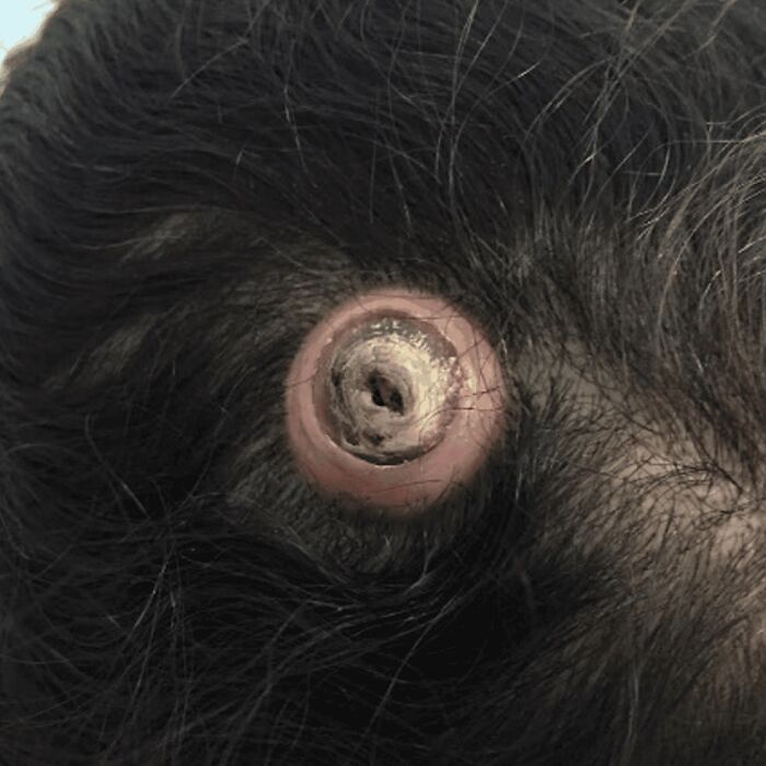
A 57-year-old female presented with a rapidly growing ellipse-shaped nodule on her scalp for two years .The mass did not regress with time and was filled with serous fluid. The patient had a medical history of diabetes and hypertension, and her family history was not significant. Physical examination revealed an oval-shaped mass with a central scar, no discharge, no fluctuation, and painless.
The gross pathological description includes a skin ellipse measuring 1.5 x 1.2 × 0.4 cm with a raised skin lesion and a central crater measuring 1.1 cm.
Microscopically, sections showed skin-abundant pilosebaceous follicles with dilated infundibulum containing lamellated keratin and lined by stratified squamous epithelium with peripheral basal cells and no cytologic atypia
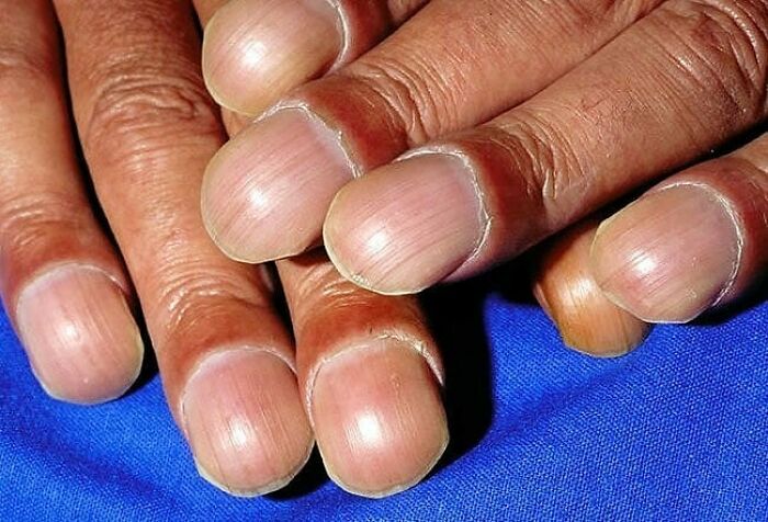
Nail clubbing is a deformity of the finger or toe nails associated with a number of diseases, mostly of the heart and lungs.
Nail clubbing occurs when the tips of the fingers enlarge and the nails curve around the fingertips, usually over the course of years. Nail clubbing is sometimes the result of low oxygen in the blood and could be a sign of various types of lung disease.
When it occurs together with joint effusions, joint pains, and abnormal skin and bone growth it is known as hypertrophic osteoarthropathy.
Clubbing is associated with lung cancer, lung infections, interstitial lung disease, cystic fibrosis, or cardiovascular disease.
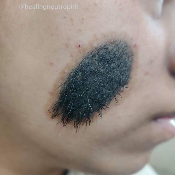
A 15-year-old female presented with a 8 cm x 7 cm, ovoid-shaped, hairy lesion on the right side of her face The palm-sized solitary lesion was present since birth and enlarged as she grew .An interview confirmed no personal or family medical history of melanoma or any form of cancer. The patient denied pain, pruritis, functional problems, or limitations in facial expression due to the lesion
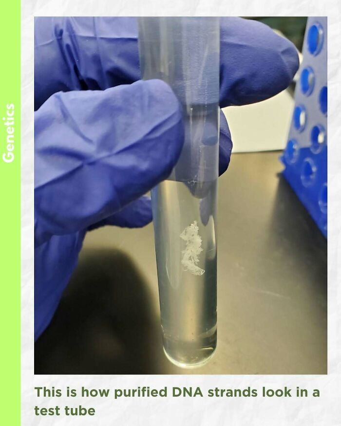
Fascinating to see purified DNA (Deoxyribonucleic acid) inside a test tube
In today’s world of DNA analysis by multiplex and real-time PCR, the importance of high-quality, purified DNA cannot be underestimated. Finding a suitable DNA isolation system to satisfy your downstream application needs is vital for the successful completion of experiments.
There are five basic steps of DNA extraction that are consistent across all the possible DNA purification chemistries: 1) disruption of the cellular structure to create a lysate, 2) separation of the soluble DNA from cell debris and other insoluble material, 3) binding the DNA of interest to a purification matrix, 4) washing proteins and other contaminants away from the matrix and 5) elution of the DNA
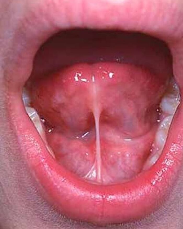
Ankyloglossia, also known as tongue-tie, is a congenital oral anomaly that may decrease the mobility of the tongue tip and is caused by an unusually short, thick lingual frenulum, a membrane connecting the underside of the tongue to the floor of the mouth. Ankyloglossia varies in degree of severity from mild cases characterized by mucous membrane bands to complete ankyloglossia whereby the tongue is tethered to the floor of the mouth.
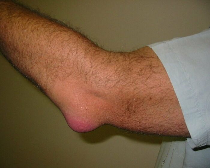
Olecranon bursitis is a condition characterized by swelling, redness, and pain at the tip of the elbow. If the underlying cause is due to an infection, fever may be present. The condition is relatively common and is one of the most frequent types of bursitis.
It usually occurs as a result of trauma or pressure to the elbow, infection, or certain medical conditions such as rheumatoid arthritis or gout.
The underlying mechanism is inflammation of the fluid filled sac between the olecranon and skin.
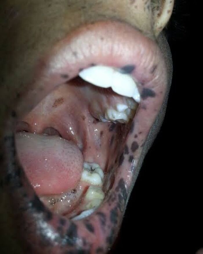
A 22 year-old male was brought to a clinic for evaluation of intermittent abdominal pain and watery diarrhea of 12 years’ duration. Over the previous 2 months, his symptoms had included vomiting and weight loss. The patient had numerous hyperpigmented macules on his lips, buccal mucosa, fingers, and toes. Computed tomography (CT) and ultrasonography showed duodenojejunal intussusception. Upper gastrointestinal (GI) endoscopy revealed multiple polyps.
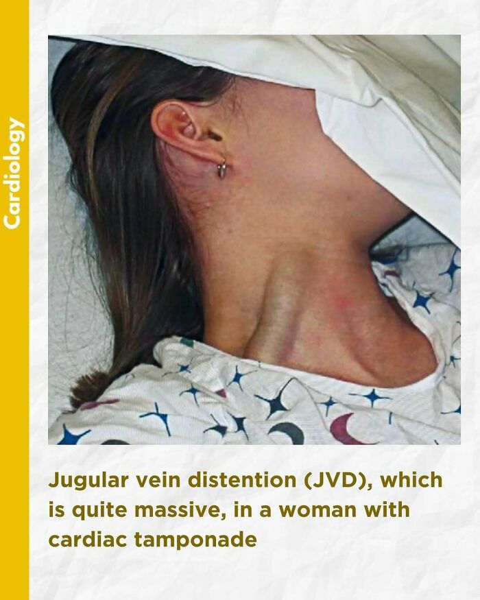
An elevated jugular venous pressure (JVP) is the classic sign of venous hypertension (e.g. right-sided heart failure). JVP elevation can be visualized as jugular venous distension, whereby the JVP is visualized at a level of the neck that is higher than normal. The jugular venous pressure is often used to assess the central venous pressure in the absence of invasive measurements (e.g. with a central venous catheter, which is a tube inserted in the neck veins).
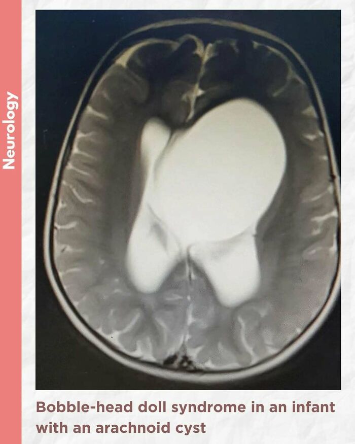
Bobble-head doll syndrome in an infant with an arachnoid cyst
Bobble-head doll syndrome is a rare neurological movement disorder in which patients, usually children around age 3, begin to bob their head and shoulders forward and back, or sometimes side-to-side, involuntarily, in a manner reminiscent of a bobble head doll. The syndrome is related to cystic lesions and swelling of the third ventricle in the brain. Symptoms of bobble-head doll syndrome are diverse but can be grouped into two categories: physical and neurological. The most common form of treatment is surgical implanting of a shunt to relieve the swelling of the brain.
A 1.5-year-old girl presented to the pediatric clinic with the chief complaints of gradual onset excessive head nodding (side-to-side movement) for 3 months. Movements increased with walking, emotions, and stress; decreased during periods of concentration; and were absent during sleep. There were no other complaints or headaches. There was no other significant history.
The child was alert, with normal cognitive function. Neurological examination was normal. Initial laboratory assessment including CBC, hepatic and renal function, and endocrine function tests were normal.
Cranial MRI demonstrated a large left-hemispheric cystic process with a midline shift, well-defined thin-walled suprasellar arachnoid cyst measuring 3 × 5 × 7 cm that obstructed the foramina of Monro, with resulting hydrocephalus ventriculomegaly. Based on the cranial MRI and symptoms, a diagnosis of a suprasellar arachnoid cyst with BHDS was made. The patient underwent endoscopic cystoventriculostomy and cystocisternostomy for the suprasellar arachnoid cyst. During the 6 months of follow-up, the head bobbing disappeared completely, and her growth was normal.
Despite the rareness of bobble-head doll syndrome, it is considered an important condition that must be investigated early to detect the cause and treated promptly to avoid potential complications.
You Might Also Like: 36 people who asked for photo edits and got amusingly unexpected results





































 No fees, cancel anytime