Get Premium
Dark mode theme is available exclusively for premium users. Learn more about the benefits of subscribing.
No fees, cancel anytime.

 Dark Mode Ad-Free Browsing Unlimited Content
 Ad-Free Browsing Unlimited Content Dark Mode
Join 1.2 million Panda readers who get the best art, memes, and fun stories every week!
Grab your lab coat, or at least pretend you have one, and prepare to zoom way, way in because we’re about to explore a world that’s been hiding right under your nose this whole time. Ever wondered what a grain of pollen looks like up close? Or why everyday objects start looking like alien landscapes the second you stick them under a microscope?
Luckily for us, curious minds have been busy capturing this hidden universe and sharing the results online. Turns out the tiniest things can be the most mind-blowing, so buckle up, because these insanely cool microscope pictures are about to make you question everything you thought you knew about the small stuff.
This post may include affiliate links.
"Mesmerizing light microscopy image from a skeleton of a diatom algae 32 to 40 million years old. Diatoms are photosynthesizing algae at the base of the marine food chain, found in almost every aquatic environment. They are single celled organisms that produce an external wall composed of silica. When they die, their silica shells accumulate on the floor of the body of water in which they live. Thick layers of these diatom shells have been fossilized into sedimentary rock called diatomite, or Diatomaceous earth!" - OCR
Microscopes open a world invisible to the bare eye, bending light, or electrons, through lenses to enlarge tiny structures. Magnification makes objects bigger, while resolution determines how clearly fine details can be seen. Motic Microscopes explains that these principles are the foundation for everything we’re about to explore.
Different microscopes reveal different aspects of the microscopic world. Light microscopes allow scientists to view different specimens, often thinly sliced or mounted in liquid, while electron microscopes require non-living, dehydrated and sometimes metal-coated samples, producing extreme detail at the cost of life. Knowing the difference helps explain why some images look so alien compared to others.
Milne Library adds that many microscope images look vividly colorful not because they naturally are, but due to staining techniques or digitally added false colors. Electron microscope images are originally black-and-white, so any color is applied afterward for contrast or visual appeal, creating almost artistic results.
These are Coccolithophores, single celled plankton. The chalk you buy to write with is synthetic, not like this at all. Natural chalk rock may contain these, but if so then I don't know where.
Further explaining this, microbiologists and histologists routinely use dyes to make transparent cells visible. Simple stains color entire cells, while differential stains, like Gram or acid-fast staining, highlight specific structures or species. For example, Gram-positive bacteria appear purple, while Gram-negative bacteria turn pink, helping scientists differentiate types of organisms under the lens.
Fascinating array of shapes and colors. I especially like the little squiggles.
Earthly Mission explains that microscopes open the door to a hidden world that looks completely different from our everyday experience. Objects that seem smooth, simple, or even boring to the naked eye suddenly reveal complex textures, intricate patterns, and entire ecosystems of microscopic life. A plain surface can turn into a rugged landscape, and something lifeless can appear full of structure and activity.
Under magnification, familiar items take on entirely new forms. Bacteria appear as rods, spheres, or spirals on surfaces we consider clean. Insects show intricate compound eyes and jointed limbs, while salt crystals reveal sharp-edged cubes. Hair shows layered textures, and fabrics expose tangled fibers often carrying dust or microbes.
I had this as my desktop background for quite a while and never ran out of things to discover.
DSS Image explains that microscopy is more than just visually striking pictures, it is essential for science, medicine, and materials research. Observations under the microscope inform medical diagnoses, drive scientific discoveries, and guide technological and quality-control advances.
In practice, hospitals and labs use microscopes to detect infections, cancer, and blood disorders by examining cells, tissues, and microbes. They help identify pathogens, monitor tumor grading, and support treatment decisions. Similarly, materials scientists use microscopes, especially SEM, to study tiny defects, cracks, and grain structures in metals and composites, improving their performance and safety.
40x total magnification, shot with an iPhone camera.
Equinecare Probiotic shares that microscopy also reveals some astonishing details: standard light microscopes can magnify up to 1,500×, turning dust into jagged landscapes, pollen into glowing miniature objects, and mold into forests of branching filaments reminiscent of alien landscapes.
I don’t shoot photos using a microscope, but a mirrorless camera with a custom built tube lens and various microscope objectives. My magnification range for images, is 10x to 40x. Sony A7R3 camera body. Tube lens using a reverse mounted Raynox DCR-150. 10x/20x/40x Mitutoyo objectives. 300 watt strobes (x2). 3 axis camera and subject positioning with sub micron resolution. 1200 pound scientific/precision granite block table on vibration isolating feet.
Looking closer at biology, human hair in cross-section shows rings and cracks like tree rings, while split ends appear frayed. Cardiac and muscle tissues form mosaic-like patterns, and red blood cells observed with electron microscopes appear as smooth, dimpled discs rather than flat circles. These details highlight the unexpected complexity in the familiar.
Finally, some microscopic animals, like loriciferans, are smaller than many single-celled organisms but still possess a brain, gut, and reproductive organs. At the atomic scale, advanced microscopy can even visualize individual atoms or layers just two atoms thick, offering a glimpse into the building blocks of matter itself.
At the heart of all these mesmerizing images, microscopy isn’t just about making tiny things look bigger, it’s about changing the way we see the world entirely. What seems ordinary at first glance can turn into something extraordinary with just a little magnification, reminding us that there’s far more detail hiding beneath the surface than we ever notice in our day-to-day lives.
Of course, not all microscopic discoveries are purely aesthetic, many play a role in science, medicine, and our understanding of life itself. However for the rest of us, they offer something just as valuable such as a fresh perspective. So the next time you glance at something as simple as a strand of hair or a grain of salt, remember you might just be looking at something far more fascinating than it seems.
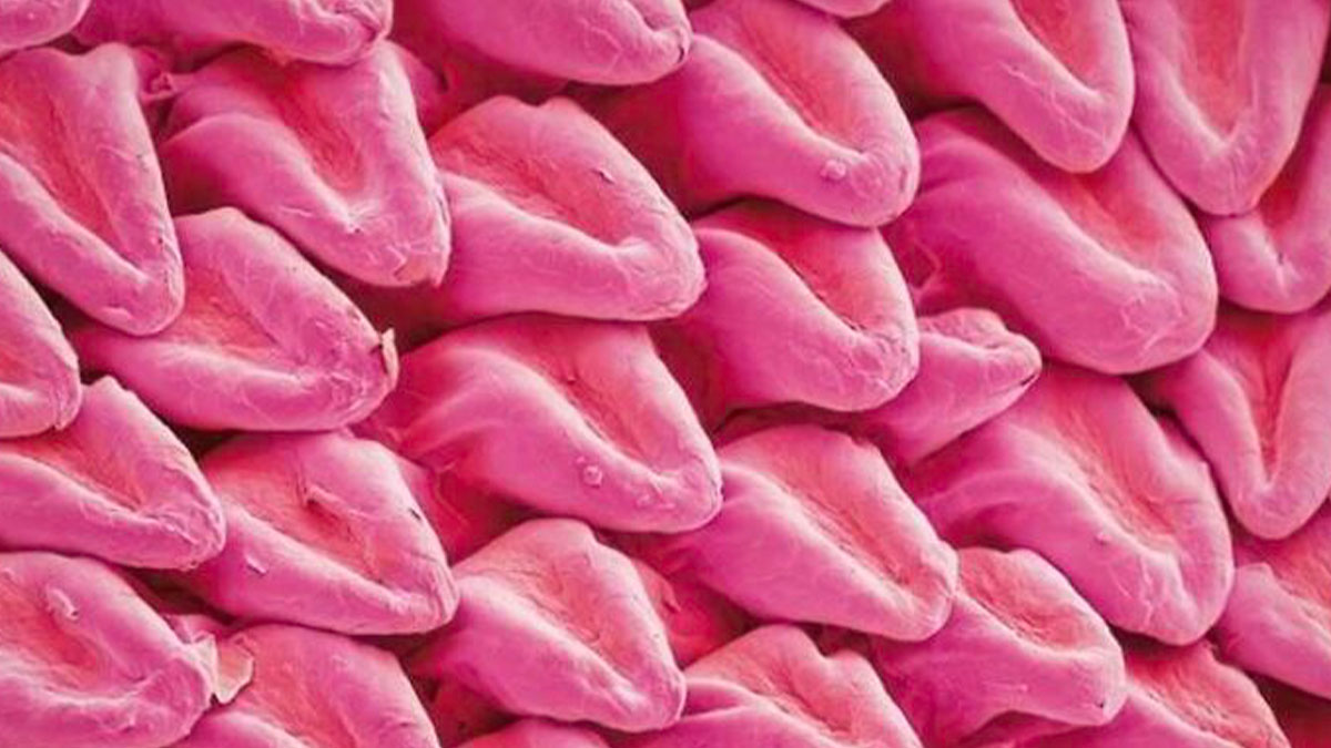
MUA Shimmer Lipstick in Rose Gold, Wet N Wild Liquid Catsuit in Berry Recognize, Tarte Lipsurgence Lipstick in Adored, ULTA Sweet and Shimmer Lip Gloss, Tarte Lipsurgence Lipstick in "Decadence", Tarte Tarteist Lip Paint.
I smeared vaseline and a surfactant (triethanolamine oleate) on glass slides. Then I performed cross-polarized optical microscopy to obtain these images. GPT enhanced the vibrancy of the blues. The blue colors result from birefringent interference. Notice the fibrous needle morphology in the vaseline compared to the lyotropic lamellae of the triethanolamine oleate.
Prions are rogue, misfolded proteins that arise in the brain and cause neurodegenerative diseases with a 100% fatality rate. They can even be transmitted between people.
I checked the Reddit post, to see what this might be. OG states that these are: "Obelia gastozooids and gonozoids. It is a thecate colonial hydrozoan."
Keep cranking that k**b down. Keep cranking... a little more... *spurt* Well done.
This type of vanadium had a beautiful rainbow-like color on it. It looks beautiful both under the microscope and in hand.


 No fees, cancel anytime


 I Investigated Under The Microscope. Also Called \"Sea Fur\"")


")


