Get Premium
Dark mode theme is available exclusively for premium users. Learn more about the benefits of subscribing.
No fees, cancel anytime.

 Dark Mode Ad-Free Browsing Unlimited Content
 Ad-Free Browsing Unlimited Content Dark Mode
Join 1.2 million Panda readers who get the best art, memes, and fun stories every week!
25submissions
1week left
Modern medicine is one of the reasons we’re able to take care of ourselves, stay healthy, and recover from illnesses that once had no cure. But getting to this point took centuries of experimentation and plenty of questionable methods along the way. You definitely wouldn’t want to be the test patient.
Unfortunately, someone had to be. Below, we’ve gathered some old medical devices that look downright creepy by today’s standards. Take a look and see if you’d have the guts to let a doctor use one on you.
Click here & follow us for more lists, facts, and stories.
This post may include affiliate links.
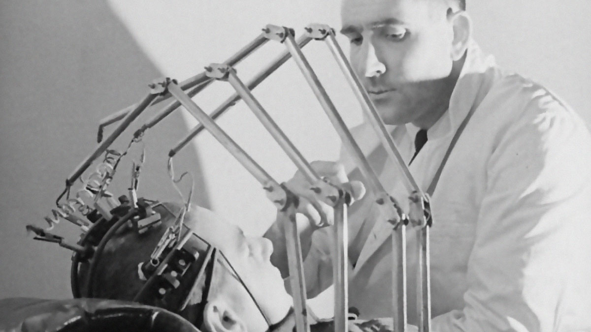
If this image doesn't scream "mad scientist laboratory," nothing does. What you're looking at is an early electroencephalograph (EEG) machine, used on a military casualty at Sutton Emergency Hospital in December 1940.
That terrifying spider-like metal cage clamped around the patient's head held multiple electrodes in place against the scalp, each one detecting the tiny electrical impulses generated by brain activity. The technology was actually groundbreaking for its time; EEGs helped doctors diagnose epilepsy, detect brain injuries, and study neurological conditions without cracking open the skull.
The Bergonic chair was used to administer electrical shocks to soldiers suffering from "shell shock," what we now recognize as PTSD. The idea was that zapping traumatized men with electricity would somehow jolt them back to psychological health, allowing them to return to the front lines.
The patient would be strapped in while a nurse operated that intimidating control panel full of dials and switches, delivering currents through electrodes attached to the body. Whether it provided any real "psychological effect" is debatable; what's certain is that men who had already endured the horrors of trench warfare were then subjected to this. Early 20th-century mental health treatment was not for the faint of heart.
"Cutting-edge cancer treatment" isn't usually synonymous with a giant ray gun pointed directly at your body. This beast of a machine at New York's Francis Delafield Hospital packed a staggering two million volts, delivering high-energy X-rays deep into tissue to target tumors that earlier, weaker machines couldn't reach.
The sheer scale of early radiation equipment is almost comical by today's standards. But machines like this represented genuine hope for cancer patients at a time when treatment options were far more limited.
This egg-shaped pod was actually custom-built for Winston Churchill. During World War II, the Prime Minister needed to travel by air for crucial meetings, but high-altitude flights posed serious risks, and Churchill wasn't exactly a young man in peak physical condition. Designer R. Graham engineered this pressurized cabin to keep Britain's leader safe and comfortable while flying above enemy territory.
Step right up, kids, and check out the machine that kept polio victims alive! This iron lung was displayed to curious visitors at Denver's Armed Forces Day celebration in 1960, a strange mix of public education and morbid fascination. The device was a negative pressure ventilator, essentially a sealed tube that rhythmically changed air pressure to force paralyzed lungs to breathe.
At the height of the polio epidemic, entire hospital wards were filled with rows of these machines, each containing a patient with only their head sticking out. Some people spent months inside; others lived in them for years or even decades. The development of the polio vaccine made iron lungs largely obsolete, which is why these kids could gawk at one as a curiosity rather than fear ending up in one themselves. Vaccines work, folks.
Nothing says "fun at the doctor's office" like strapping a child to a plastic horse and blasting them with radiation. The Roentgen Steed (named after Wilhelm Röntgen, the discoverer of X-rays) was designed to solve a real problem: getting squirmy kids to sit still long enough for a clear chest X-ray. The solution? Mount them on a little toy horse with restraints to keep them in position while the equipment does its work.
Nothing like having your legs vacuum-sealed like leftover pot roast. This contraption encased the patient's lower limbs in large cylindrical chambers that used alternating pressure (essentially squeezing and releasing) to help push blood through the extremities.
The treatment targeted patients with poor circulation, particularly those suffering from peripheral vascular disease or conditions that left blood pooling sluggishly in the legs.
The Electric Bath was an early form of light therapy, essentially a coffin-shaped chamber lined with electric bulbs that bathed the patient in intense illumination. Think of it as the great-great-grandfather of the modern tanning bed, but with more "medical legitimacy" and less concern about UV damage. At the time, light therapy was believed to treat everything from skin conditions to fatigue to depression, and honestly, they weren't entirely wrong about some of it.
Developed by Dutch physiologist Willem Einthoven in the early 1900s, the first electrocardiogram machine detected the heart's electrical impulses using saline-filled containers as conductors, since electrodes hadn't quite been perfected yet.
The apparatus required an entire room's worth of equipment just to produce a single reading of cardiac activity. Einthoven won the Nobel Prize in 1924 for his invention. Today's EKGs involve a few small stickers and take about ten seconds; back then, you showed up and put your feet in buckets. Medicine has come a long way.
When your microscope is taller than you are, you know you're doing serious science. This towering column of metal and dials is an early electron microscope, a device that used beams of electrons instead of light to magnify specimens at levels traditional microscopes could never achieve, revealing the hidden world of viruses, cell structures, and molecular details.
The technology was revolutionary when it emerged in the 1930s, allowing scientists to see things thousands of times smaller than what optical microscopes could manage. Of course, operating one required an entire room, a wall of controls, and presumably a very patient researcher.
Here, a doctor is casually observing a live X-ray of his patient's ribcage displayed on a fluoroscopic screen like some kind of ghostly window into the human body. At Professor Menard's radiology department in Paris, this was state-of-the-art diagnostic technology, allowing physicians to peer inside the chest without making a single incision.
Here's the terrifying part: notice what's missing? No lead aprons, no protective barriers, nothing between the doctor and a constant stream of radiation. Early radiologists had no idea how dangerous prolonged X-ray exposure was; many pioneers in the field developed radiation burns, cancers, and other horrific ailments. Some even lost limbs. The technology was revolutionary, but the price paid by those first practitioners was steep.
Finally, an iron lung you could take home! This compact chest respirator was a major upgrade from the massive full-body tubes that kept polio patients trapped in hospital wards. Instead of sealing a person inside a giant metal cylinder, this device simply clamped onto the chest and used an external pump to do the breathing work.
The design allowed patients to recuperate in their own homes, a revolutionary concept when polio wards were overflowing and the psychological toll of living inside a machine was immense. By this point in 1955, the Salk vaccine was already being distributed, and polio's reign was finally coming to an end.
It looks like a scene from a mad scientist's lair, but this was simply another day at the office for Nurse Nataly Nyepomnachai. That tangle of wires erupting from the patient's skull is just standard EEG equipment, designed to measure electrical activity in the brain.
The nurse carefully manages each electrode while the patient sits remarkably still beneath the chaos. EEG technology was invaluable for diagnosing conditions like epilepsy and studying brain function, but here it was used to test the brain activity of a cosmonaut preparing to go to space.
This advertisement for the Oxyoline Apparatus shows off an early oxygen delivery system. The large wooden cabinet housed the equipment needed to pump supplemental oxygen through the hose the nurse is cheerfully holding up.
Manufactured by the Neel-Armstrong Company out of Akron, Ohio, the device was designed for patients suffering from respiratory distress, pneumonia, or other conditions where a little extra oxygen could mean the difference between life and the grave.
Sweet dreams are made of... whatever gas is coming out of that contraption. This early anesthesia machine features an elaborate system of gears, cranks, and tubes connected to a mask strapped firmly over the patient's face, operated by a mustachioed physician who looks like he absolutely knows what he's doing. Hopefully.
Before machines like this, surgery was either agonizingly painful or relied on crude methods like alcohol, opium, or simply knocking the patient unconscious. The development of controlled anesthetic delivery was revolutionary, finally allowing doctors to perform complex procedures while patients slept peacefully instead of screaming. Of course, "controlled" is a relative term here; that hand-cranked apparatus required constant manual adjustment, and dosing was more art than science. One wrong turn of the dial and your patient either woke up mid-surgery or didn't wake up at all.
This nurse operates an early X-ray machine, carefully positioning the articulated metal arm and tube to capture an image of her patient. Early radiological workers often operated these machines dozens of times a day without any understanding of cumulative exposure. Today's X-ray technicians step behind protective walls and wear monitoring badges; back then, everyone just stood in the radiation together and hoped for the best.
Dr. G.H. Byford stands beneath a rotating optokinetic drum wearing a contact lens with a tiny lamp cemented directly onto it—allowing researchers to track exactly how his eyes moved in response to visual stimuli spinning around him.
The experiment investigated reflex eye movements and their connection to visual illusions, crucial information for understanding why pilots sometimes become dangerously disoriented during flight. The RAF Institute of Aviation Medicine at Farnborough was constantly dreaming up contraptions like this to study human perception under extreme conditions.
When regular eyeball diagrams just won't cut it, you build a pair the size of beach balls. Cpl. Charles F. Morris of Bristow, Oklahoma, poses proudly between these massive motorized eye models used to teach aviation medical examiners at Randolph Field how ocular muscles actually work.
The oversized specimens lit up and moved via small internal motors, allowing large classrooms to observe eye movements in real time during lectures. Understanding how pilots' eyes function under stress was crucial for aviation medicine, and these gloriously creepy teaching tools made sure every student in the room could see exactly what was happening.
Just a scientist and his transparent best friend, casually discussing the effects of nuclear radiation. "Plastic Man" was a life-sized human phantom made of clear plastic with a full anatomical skeleton and was a crucial research tool used to simulate how radiation travels through and affects the human body.
By embedding dosimeters and sensors throughout the phantom, scientists could measure exactly how much radiation reached different organs and tissues without, you know, irradiating actual people. This was essential work during the Cold War era, when understanding radiation exposure was a matter of national security and public health.
And you thought wearing a paper mask to the grocery store was inconvenient. This woman is casually reading while connected to an early respiratory filtration device during the devastating 1918-1919 Spanish Flu pandemic.
With no vaccines and limited understanding of how viruses spread, people got creative with protective measures. While most simply wore cloth masks (sound familiar?), some contraptions like this attempted to filter or purify the air before it reached the wearer's lungs. Whether this particular device actually worked is debatable, but you have to admire the commitment.
Here's a creative way to track your blood flow: inject radioactive iodine into one wrist and see how long it takes to show up at the other. This diagnostic technique measured circulation time by detecting when the radioactive tracer completed its journey through the cardiovascular system, providing valuable information for cancer diagnosis and other conditions.
Abnormal circulation times could indicate tumors, heart problems, or blockages affecting blood flow. It was an elegant solution for its era as it was using the body's own circulatory system as a delivery route and radioactivity as a tracking beacon. The patient just had to lie there, relax, and try not to think too hard about the radioactive material coursing through their veins.
When in doubt, burn it off. This nurse operates an electrocautery machine while treating lesions on her patient's forearm. Electrocautery uses electrical current to heat a metal probe, essentially creating a precise burning tool for removing unwanted tissue like warts, moles, or small growths, and for sealing blood vessels to control bleeding during procedures.
Welcome to the Sun Ray Department, where the cure for whatever ails you is being blasted with artificial light from multiple angles. Light therapy, or heliotherapy, was wildly popular during this era, and doctors believed that concentrated doses of ultraviolet light could treat everything from tuberculosis to rickets to skin conditions and general fatigue.
This experiment was part of ongoing research into reflex eye movements and visual illusions at the RAF Institute of Aviation Medicine. Understanding how eyes behave involuntarily was critical for aviation safety; pilots experiencing spatial disorientation could easily misread their instruments or lose their sense of direction, with very serious consequences.
By tracking exactly how the eye responds to various stimuli, researchers could better understand why certain illusions occur and how to train pilots to overcome them.
The reality of cancer treatment for much of the 20th century was much scarier than today (if that was even possible). This patient at the Tirana Cancer Institute receives cobalt-60 therapy, a form of radiation treatment that used the radioactive isotope to bombard tumors with gamma rays in hopes of destroying malignant cells.
The sheer size of these cobalt units was intimidating by design; the bulky housing was necessary to shield the intensely radioactive source material when not in use. During treatment, a small aperture would open to release a precisely targeted beam at the patient's tumor. It wasn't comfortable, it wasn't quick, and the side effects could be brutal, but for many cancer patients, it represented the best chance at survival.


 No fees, cancel anytime