Self Reflected: A Snapshot Of The Human Brain Captured By Reflective Microetching
Combining the circuit dynamics of ~500,000 hand drawn neurons, MRI data from the human brain, 1750 sheets of 22 karat gold leaf, and a custom LED light display comprising over 144 lights – Self Reflected provides its viewers with an unprecedented artistic glimpse into the brain activity that drives us all.
Created by artist/neuroscientist Dr. Greg Dunn and artist/applied physicist Dr. Brian Edwards, Self Reflected is designed to depict what our brains are doing while we take in this body of art. The piece is a massive installation representing a thin sagittal slice of human brain (the end result being 22 times its actual scale), chosen at a location that showcases the diverse neuronal networking that comprises our brain.
Dr. Dunn and his team utilized a technique called reflective microetching – a technique developed by Drs. Dunn and Edwards specifically to allow dynamic and revolutionary control over their images – to create this elaborate installment. Their goal is to use rigorous neuroscience and creative engineering to provide observers with a deeper understanding of our human consciousness. To learn more about reflective microetching and see other pieces created by this inventive method, check out http://www.gregadunn.com/category/microetchings/.
Self Reflected is on permanent exhibition at the Franklin Institute in Philadelphia, and was funded by the National Science Foundation. Microetchings and prints of these beautiful microetchings as well as other projects by Dr. Dunn are available at http://www.gregadunn.com, where you can also watch Self Reflected (and videos of other microetchings) in action. For more information on how Self Reflected was created, and the artistic and scientific basis behind it, visit http://www.gregadunn.com/self-reflected/.
More info: gregadunn.com
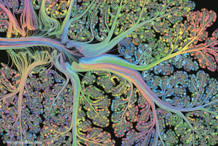
The laminar structure of the cerebellum, a region involved in movement and proprioception (calculating where your body is in space). Self Reflected (detail), 22K gilded microetching, 96″ X 130″, 2014-2016, Greg Dunn and Brian Edwards.

The entire Self Reflected microetching under white light– (photo by Greg Dunn and Will Drinker). Self Reflected (detail), 22K gilded microetching, 96″ X 130″, 2014-2016, Greg Dunn and Brian Edwards.

The visual cortex, the region located at the back of the brain that processes visual information. Self Reflected (detail), 22K gilded microetching, 96″ X 130″, 2014-2016, Greg Dunn and Brian Edwards.

The brainstem and cerebellum, regions that control basic body and motor functions – (photo by Greg Dunn and Will Drinker). Self Reflected (detail), 22K gilded microetching, 96″ X 130″, 2014-2016, Greg Dunn and Brian Edwards.

The parietal gyrus where movement and vision are integrated– (photo by Greg Dunn and Will Drinker). Self Reflected (detail), 22K gilded microetching, 96″ X 130″, 2014-2016, Greg Dunn and Brian Edwards.

The motor and parietal cortex, regions involved in movement and sensation, respectively– (photo by Greg Dunn and Will Drinker). Self Reflected (detail), 22K gilded microetching, 96″ X 130″, 2014-2016, Greg Dunn and Brian Edwards.

213views
Share on Facebook

17
3