Get Premium
Dark mode theme is available exclusively for premium users. Learn more about the benefits of subscribing.
No fees, cancel anytime.

 Dark Mode Ad-Free Browsing Unlimited Content
 Ad-Free Browsing Unlimited Content Dark Mode
Join 1.2 million Panda readers who get the best art, memes, and fun stories every week!
14submissions
1week left
Despite the fact that we quite literally embody our own bodies, they can still be a source of horror in the right (or wrong) circumstances. It’s one of the reasons roughly 4% of US medical students drop out, not due to poor grades but the realization that this maybe isn’t for them.
We’ve gathered some unsettling, creepy but perhaps still interesting pictures about the unusual things that can go on in and outside of the human body. Be warned, some of these images can be unpleasant. Otherwise, settle in as you scroll through, upvote your favorites and be sure to share your thoughts in the comments down below.
Click here & follow us for more lists, facts, and stories.
This post may include affiliate links.
Scoliosis is characterized by a lateral deviation and rotational deformity of the spine, resulting in an abnormal sideways curvature. It can manifest in different regions of the vertebral column and exhibit varying degrees of severity.
This 13-year-old’s scoliosis was progressing so rapidly that major spinal surgery was her only treatment option. In just over six months, her curve progressed from what was initially 49-degree to a 99-degree curve. The girl now has a combination of titanium rods and screws around her spine. Luckily she fully recovered and got back to her normal activities.
While scoliosis can be caused by conditions such as cerebral palsy and muscular dystrophy, the cause of most scoliosis is unknown. About 3% of adolescents have scoliosis.
Treatment depends on the degree of curve, location, and cause. Minor curves may simply be watched periodically. Management options may involve close observation, utilization of orthotic devices (e.g., braces) for stabilization, or, in severe cases, surgical intervention aimed at rectifying the curvature and achieving spinal stability. The brace must be fitted to the person and used daily until growing stops.
Surgery is usually recommended by orthopedists for curves with a high likelihood of progression (i.e., greater than 45 to 50° of magnitude), curves that would be cosmetically unacceptable as an adult, curves in people with spina bifida and cerebral palsy that interfere with sitting and care, and curves that affect physiological functions such as breathing. To completely straighten a scoliotic spine is usually impossible, but for the most part, significant corrections are achieved.
Credit: Isabel Dayman, Mobile.abc.net.au
Possessing menacing eyes and devilish grins, you would be forgiven for assuming these were just aliens from a Hollywood sci-fi blockbuster.
These images were circulating on social media apps and while some suggest they have been traumatised by viewing such discomforting images, others seem to find them comical.
Some users even believed the fetus was an extra-terrestrial being.
Yet, believe it or not, they are not fake.
Instead, they are genuine MRI scans of human babies in the womb.
MRI scans are different to ultrasounds. Parents are not regularly offered MRI throughout their pregnancy and will typically only have the scan if there is a concern for the child's growth and development.
For example, they can help define and detect neck, thoracic, abdominal and spinal malformations in fetuses.
When used during pregnancy, however, MRIs can produce a very life-like image of their baby.
The detailed black and white images burst the bubble of many parents who blindly believe their tiny tot is going to be adorable through and through.
One user said MRIs are discouraged during pregnancy because 'people would realise they're incubating nightmare demons and would be rightfully terrified'.
We can confirm these images are authentic and real.
MRI uses magnetic fields and radio waves to produce detailed images of the inside of the body.
The eyes and brain have high levels of 'signal' — a radio wave — which causes them to appear brighter and stand out on the scan.
Other parts of the body give off lower levels, and therefore appear darker.
A 67-year-old woman scheduled for routine cataract surgery thought it was just dry eye and old age causing her discomfort, she told her surgeons.
But what doctors found to be the real cause of her discomfort was much more concerning: 27 disposable contact lenses, stuck in the woman's right upper eyelid.
An anesthetist at the hospital was beginning to numb her eye for surgery when he found the first cluster of contacts.
He put a speculum into the eye to hold the eye open as he put the anesthetic in, when he noticed a blue foreign body emerging from the top eyelid.
That mass was a clump of 17 lenses (bottom picture).
On closer inspection, 10 more lenses were discovered floating around loose.
The lenses were clumped together in a “blueish mass” and were bound together by mucus.
The woman had been wearing monthly disposable contact lenses for 35 years, but it's unclear how long they had been gathering in her eye. Sometimes, she told the surgeons, when she would try to remove a contact from that eye, she couldn't find it.
The patient had just figured she'd dropped it somewhere, but it was actually getting stuck in her eye with the others.
Two weeks after removing the lenses her eyes felt a lot more comfortable.
Source - the British Medical Journal.
The human body exists as a walking contradiction, a masterpiece of biological engineering that simultaneously serves as a source of profound existential dread. This dualistic relationship stems from a deeply rooted psychological phenomenon known as the uncanny valley, where something that is almost, but not quite, human triggers a sense of unease or even revulsion. When we look at a medical cross-section or a high-definition image of a cellular structure, we are forced to confront the mechanical reality of our existence.
We like to think of ourselves as coherent personalities with dreams and memories, but medical imagery reminds us that we are also a complex assembly of pulsing valves, electrical signals, and strange, wet textures. This confrontation with our own biological materiality is where the horror begins, yet it is exactly this complexity that keeps us clicking through gallery after gallery of anatomical wonders.
Scientists last month published an unprecedented case, where they found and extracted a live parasitic worm from the brain of a 64-year-old Australian woman.
The neurosurgeon found and removed the parasite with forceps during a biopsy, from within the lesion shown on the MRI (light gray area).
"I used tumor-holding forceps and lifted out something that I definitely was not expecting: a linear, squiggling line, and my junior doctor said, 'is that an artery?', because that's what it looked like. And I said, 'it's not an artery, we're nowhere near any artery!' And I noticed it was moving and I went, 'just get it out of my forceps!' So we rapidly put it in a pathology pot, and it was a vigorously wriggling worm."
Symptomatically, weeks of abdominal pain and diarrhea led to night sweats and a dry cough, but evolved towards problems like forgetfulness and depression, presumably as the worm׳s activities kept affecting different parts of the brain.
The worm was some 8 centimeters (just over 3 inches) long and is a rare parasite called Ophidascaris Robertsi. This is a type of Roundworm (Helminth). This roundworm usually lives in a Carpet Python. The eggs of the worm are around the snakes’ faecal droppings, which infect the grass. This grass containing the eggs, are eaten by small mammals, who are then eaten by the Carpet Python. This is how this worm gets cycled between its two hosts. This woman became an 'accidental host'. She lived near the carpet python habitat and while foraging the native vegetation for cooking, she ingested the worm eggs. The worm developed in her intestine and travelled via blood circulation to her brain.
In response, the body produces inflammation around the worm and as a result, an area of inflammatory tissue or granuloma develops in the brain. Depending upon the affected location, it can cause a multitude of symptoms such as pressure symptoms - headache, vomiting, visual blurring, confusion, altered sensorium, cognitive symptoms - forgetfulness, problems in understanding, calculations, disorientation, seizures and epilepsy - due to irritation of the brain by inflammatory tissue.
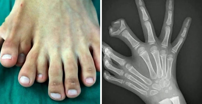
This very specific clinical finding is associated with neurofibromatosis type 1 (NF1), also called von Recklinghausen's disease, a rare genetic disorder characterized by the development of multiple noncancerous (benign) tumors of nerves and skin (neurofibromas) and areas of abnormally decreased or increased coloration (hypo- or hyperpigmentation) of the skin. It is one of the most common genetic disorders and affects 1 in 3,500. Adults develop neurofibromas, which are noncancerous (benign) peripheral nerve sheath tumors that are usually located on or just under the skin. Many have also multiple café-au-lait spots, which are flat patches on the skin that are darker than the surrounding area. NF-1 is caused by a mutation of a gene on the long arm of chromosome 17 which encodes a protein known as neurofibromin, which is a negative regulator of the Ras oncogene signal transduction pathway. When Ras isn’t regulated, it is overexpressed. These are a family of proteins that are involved in cellular signal transduction. A cascade effect occurs when ras is “switched on” by incoming signals, leading to activation of other proteins, which, in turn, activate genes responsible for cell growth and differentiation, hence leading to neurofibromas. Due to their benign nature, neurofibromas should be surgically excised only when symptomatic.
Our fascination is partly fueled by the negativity bias, a survival mechanism that compels us to pay more attention to things that could potentially harm us than things that are pleasant. From an evolutionary perspective, being repulsed by signs of disease or physical trauma was a way to keep our ancestors safe from infection.
They are present in neonates or infants but they may also be present prenatally and at birth they may become large enough to cause obstructed labor. The swelling is translucent, usually bilateral, soft and partially compressible and it's size increases when the child cries or coughs. The swelling can sometimes become so large that it causes respiratory problems and feeding difficulties which makes it difficult for the child to thrive. The cyst can also become infected if not treated. The only definitive treatment is complete excision of the cyst when it is at an early stage. However, it is essential that no residual tissue is left behind otherwise it may recur
When we see a medical photo of a rare pathology or a distorted bone structure, our brains are essentially running a diagnostic scan, assessing the threat and learning what to avoid. However, because we are viewing these images from the safety of a screen, that primal fear transforms into a form of morbid curiosity.
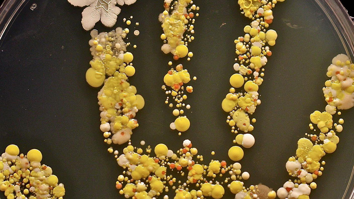
The boy came in from playing outside, and his mom decided to put his hand print inside a large Petri dish, incubated it for two days, and ended up with a colorful germ garden!
Consider for a moment what you've touched today. After all, our hands do all sorts of things for us – they open doors, handle money at the wet market, hold on to poles on public buses and trains, all the time while grasping our phones. They often also act as the barrier between our coughs and the rest of the world.
Up to 80% of all infections are transmitted by hands.
The colony on the hand would consist of various bacteria, potentially including both gram-positive and gram-negative species. These bacteria could belong to different genera and species, such as Staphylococcus, Streptococcus, or Escherichia coli, among others. The colony may exhibit different morphological characteristics, such as size, shape, and coloration, depending on the specific bacteria present.
Additionally, the colony might demonstrate characteristics indicative of bacterial growth, such as a visible texture or elevation. It could display features like smoothness, roughness, or irregularity. The Petri dish would provide a suitable environment for these bacteria to multiply, as it would contain a culture medium that supports their growth and sustenance.
We are allowed to gaze upon the "forbidden" interior of the species without any actual danger, satisfying a voyeuristic urge that has existed since the first public dissections in the Renaissance. The aesthetic of the internal body also plays a massive role in our fixation. There is an undeniable, albeit alien, beauty in the fractal patterns of the bronchial tubes or the vibrant, dyed landscapes of a histological slide.
You Might Also Like: 26 Absurd Job Requirements That Are Exactly Why “No One Wants To Work Anymore”


 No fees, cancel anytime


")


