when you think you have seen everything these German guys come with pictures out of this world. These are fantastic microphotographs taken under the Scanning Electron Microscope. Nicole Ottawa (Biologist) and Oliver Meckes (Photographer), joined their expertise to found Eye of Science since 1995 to delight us with fantastic never-seen-before images of organisms, microorganisms and thousands of other stuff that seem to come out of this world.
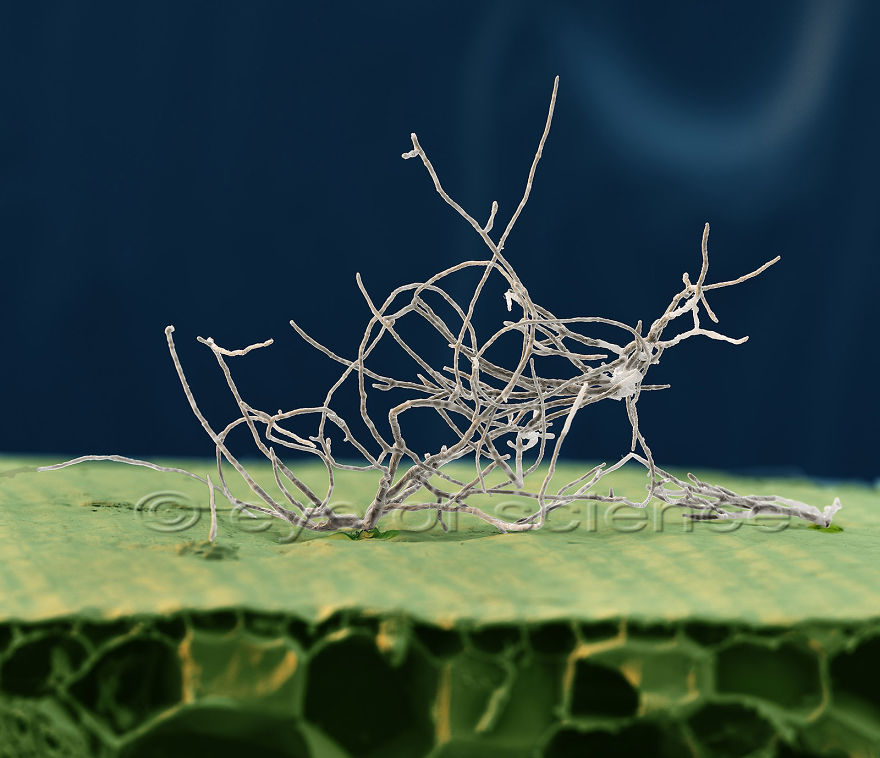
Recently, we worked together to obtain pictures of Fusarium spp. the pathogen that causes the Panama disease of bananas. the result was impressive (you can see one of our pictures here), I decided to explore a bit more of their work and I can say that I will never regret.
Try to guess before you read the answer.
To see more of their work visit Eyeofscience.de
to see more of the banana fungus visit @ferchucky
enjoy
More info: Instagram
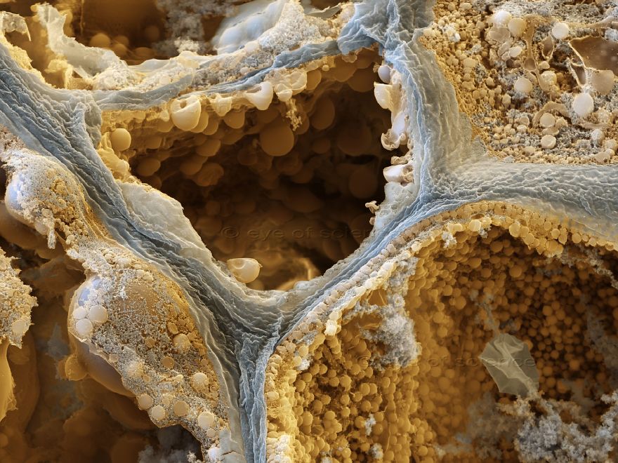
This scanning electron micrograph shows the fungus Fusarium oyxsporum f.sp. cubense, causal agent of the Panama disease of bananas. Cavendish bananas are resistant to the Race 1 but extremely susceptible to the Tropical Race 4 (TR4). TR4 is spreading and has been recently reported for the first time in the American continent where major banana producers are located. TR4 threatens the global banana production. The infected banana stem were provided by the researcher Fernando Garcia-Bastidas who does research at Keygene and Wageningen University. The image illustrates the fungus emerging from the pseudostem of a infected cavendish plant. Magnification 600:1 (when 15cms wide).
Image credits: www.instagram.com
Image credits: www.eyeofscience.de
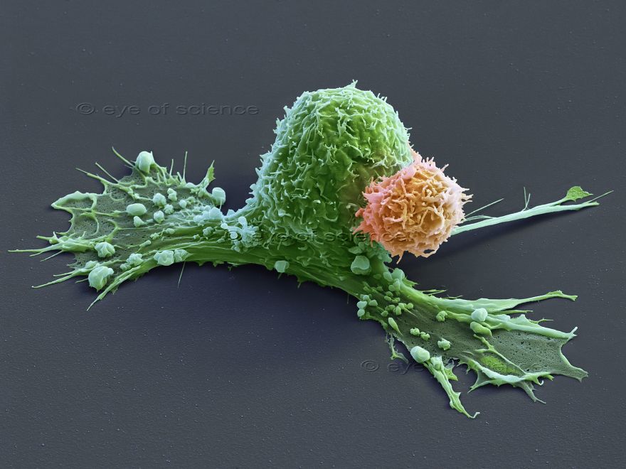
Cancer Therapy A cancer cell is attacked by a chimeric antigene receptor cell (CAR cell). Scanning Electron Microscope, 3600:1 (when 15cms wide)
Image credits: www.eyeofscience.de
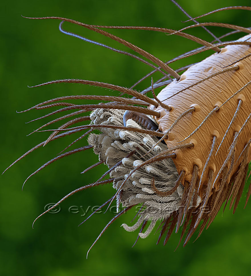
Spider feet
Image credits: www.instagram.com
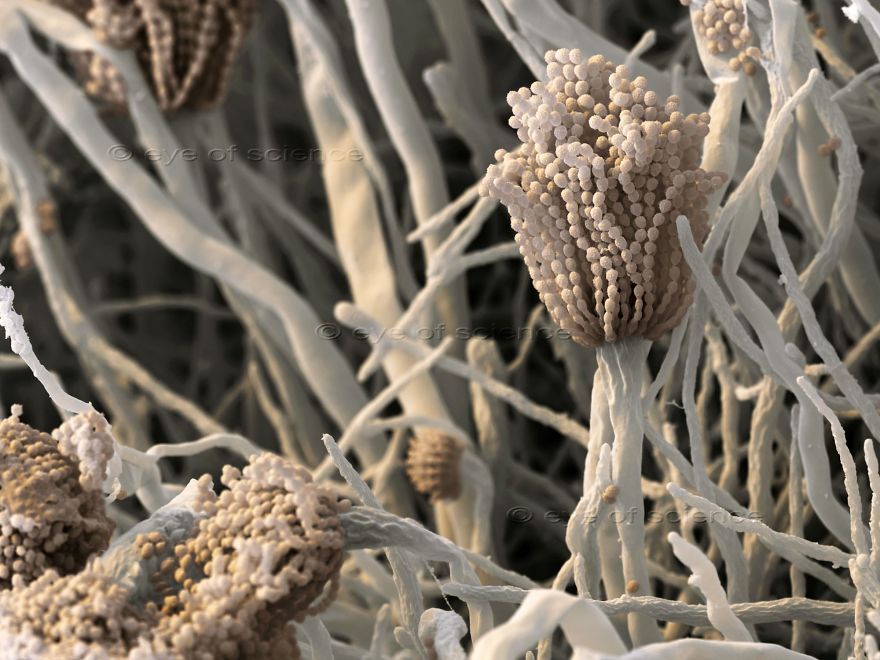
Aspergillus fumigatus Aspergillus fumigatus is a common fungus that may cause infections in immunodeficient people if the spores are inhaled. Scanning Electron Microscope, 800:1 (when 15cms wide)
Image credits: www.instagram.com
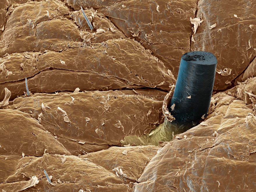
Hair Skin surface with a beard hair that has been cut one day before. Scanning Electron Microscope, 120:1 (when 15cms wide)
Image credits: www.instagram.com
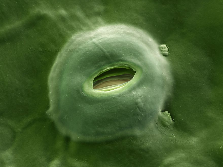
Stomata Stomata are the openings for breathing in plant leaves. There the gas exchange CO2 – O2 takes place. Scanning Electron Microscope, 4000:1 (when 15cms wide)
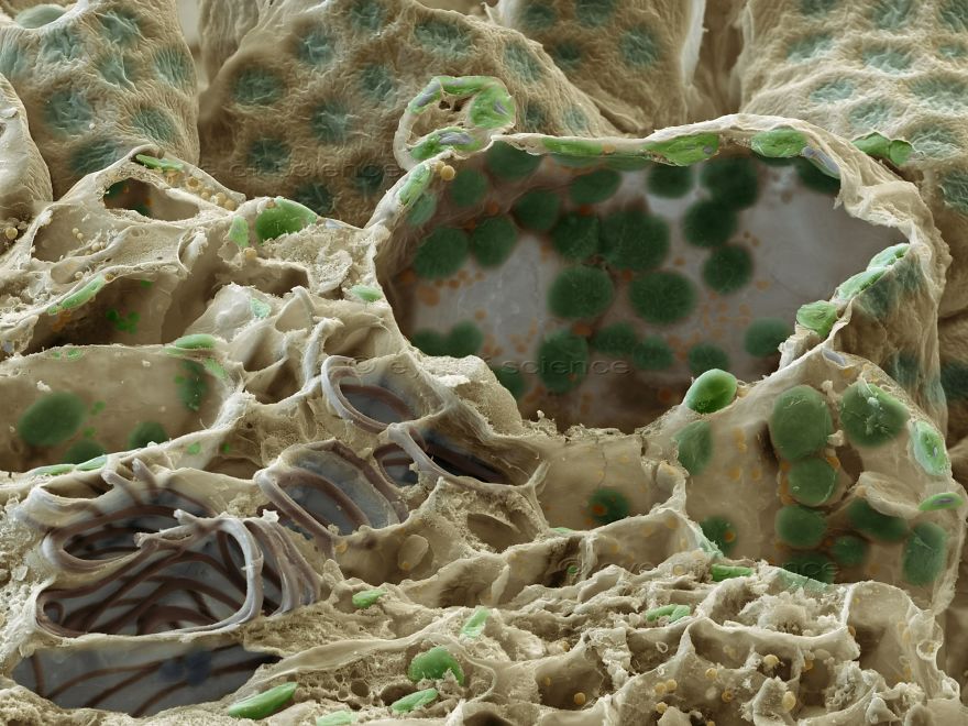
Leaf Vascular bundles (lower left) and chloroplasts (green) can be seen in this freeze fraction of a leaf. Scanning Electron Microscope, 1000:1 (when 15cms wide)
Coffee Bean Freeze fraction of a Coffee bean showing the content of the cells. Scanning Electron Microscope, 5000:1 (when 15cms wide)
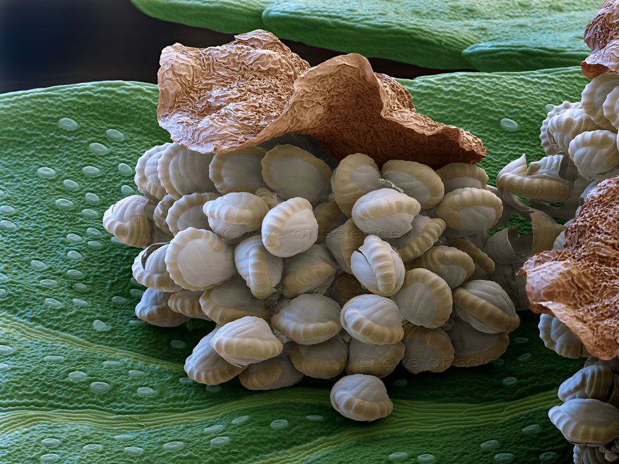
Fern Open spore capsule of a fern plant, the spores are visible. Scanning Electron Microscope, 80:1 (when 15cms wide)
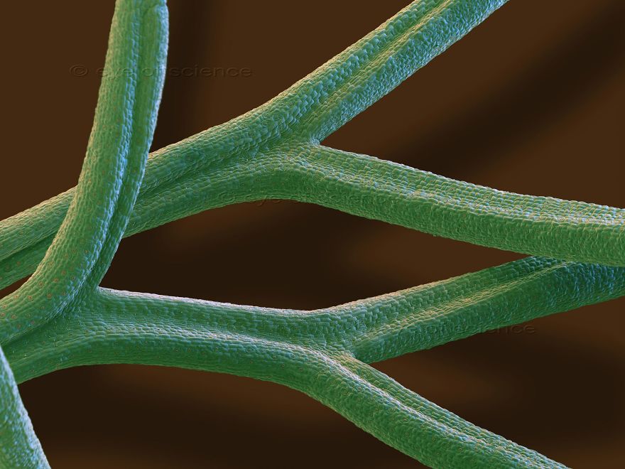
Fennel Fennel leaves with tiny stomata (brown spots) all over. Scanning Electron Microscope, 50:1 (when 15cms wide)
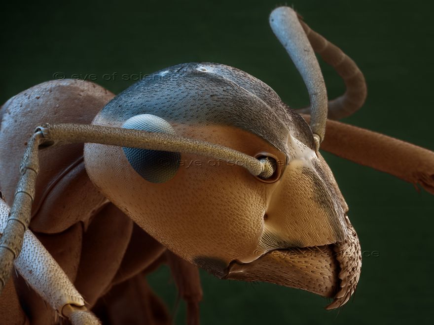
Ant Portrait of an ant with feeler, compound eyes and mandibles. Scanning Electron Microscope, 45:1 (when 15cms wide)
Image credits: www.instagram.com
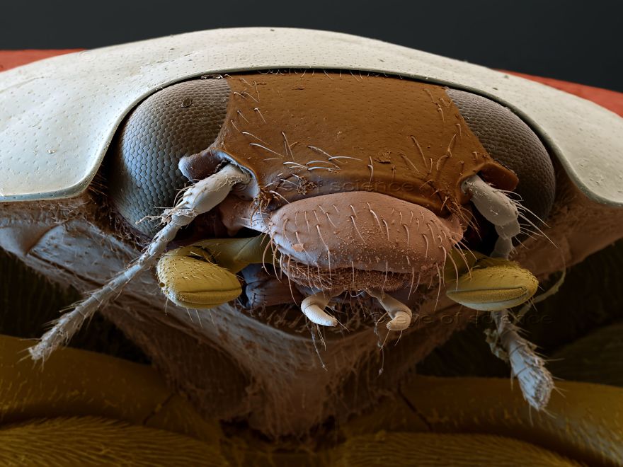
Ladybug Portrait of an asian ladybug (Harmonia axyridis). Scanning Electron Microscope, 46:1 (when 15cms wide)
Image credits: www.instagram.com
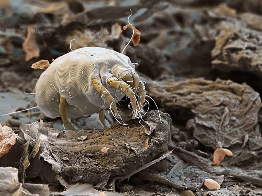
Dust Mite Dermatophagoides house dust mite between skin scales. Scanning Electron Microscope, 400:1 (when 15cms wide
Image credits: www.instagram.com
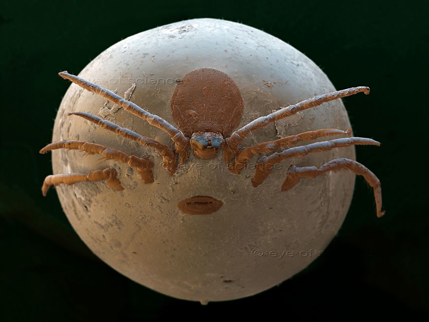
Wood Tick Fully soaked Ixodes ricinus tick. Scanning Electron Microscope, 18:1 (when 15cms wide)
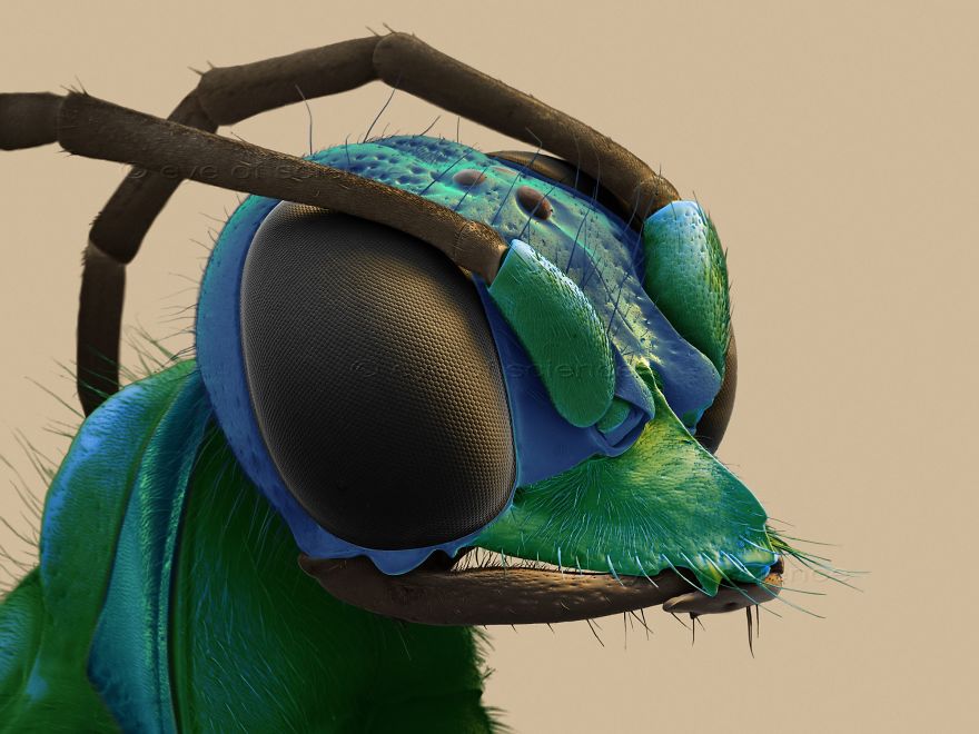
Jewel Wasp The emerald cockroach wasp or jewel wasp (Ampulex compressa) is a solitary wasp laying its eggs on cockroaches. Scanning Electron Microscope, 25:1 (when 15cms wide)
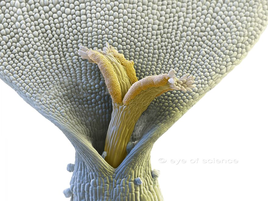
Chamomilla Single blossom of a Chamomile flower. Scanning Electron Microscope, 170:1 (when 15cms wide)
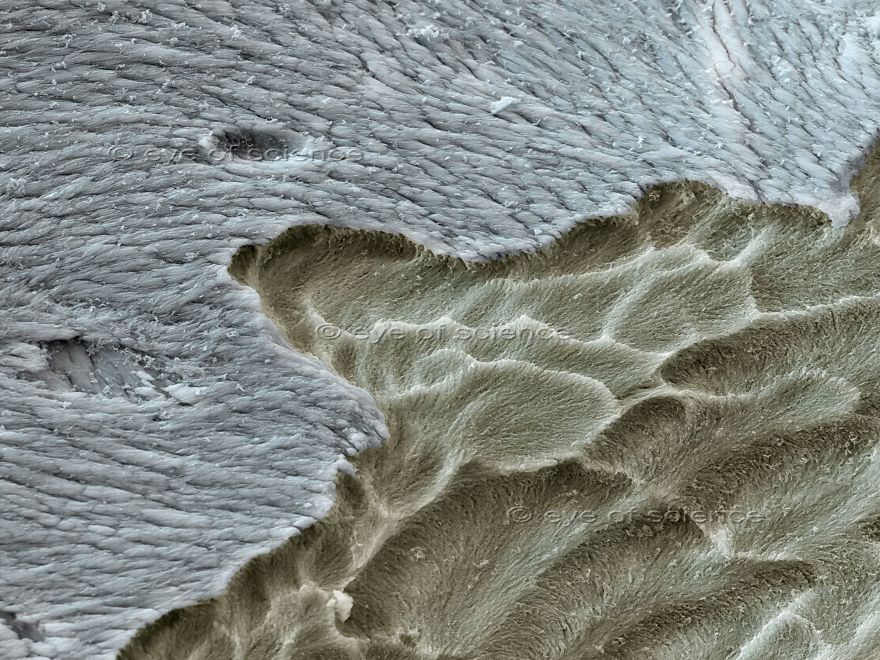
Osteoporosis Surface of a bone with osteoporosis (beige). Scanning Electron Microscope, 1000:1 (when 15cms wide)
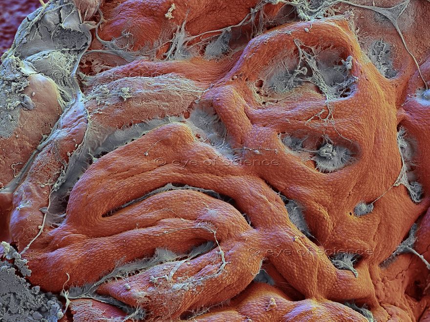
Stomach Surface of the mucous membrane of the stomach (gaster) . Scanning Electron Microscope, 230:1 (when 15cms wide)
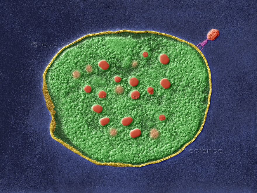
Bacteriophage Ultra thin section of a Escherichia coli bacterium infected with T-Phages. Transmission Electron Microscope, 90000:1 (when 15cms wide)
1Kviews
Share on Facebook


13
3