Looking at the World through a Microscope (Part II)
Did you enjoy looking at the World through a Microscope (Part I)? Most of the pictures on this post series were taken with an electron microscope and I completely forgot to explain what it is and how it works. Panda is very sorry for that. So, here’s some info about the different types of microscopes and how they work.
You may have seen a typical light microscope (a.k.a. optical microscope) in a science lab or at school before. If you looked down the world’s most powerful light microscope, you’d be able to distinguish individual objects that are around 200 nanometres apart — roughly 1/500th the width of a human hair. But objects closer together than that would just merge into one. This is because the wavelength of visible light is longer than 200 nanometres.
However, the difference between light microscopes and electron microscopes is the latter uses beams of electrons instead of light. The wavelength of these electron beams is much shorter, allowing scientists to see structures as small as 1 nanometre (1 millionth of a millimeter).
There are two types of electron microscopes, creating different types of images for different purposes. Transmission electron microscopes (TEM) fire beams of electrons straight through prepared samples of cells, picking up fine details of the tiny structures within. This technique allows scientists to see what’s going on deep within cells under a microscope — effectively a “molecule’s eye view”. Scanning electron microscopes (SEM) are slightly different. Instead of firing beams straight through a sample, the beams are angled so they bounce off the cell surface, providing detailed three-dimensional electron microscope images.
But we are here not for the in-depth scientific study, right? Let’s jump straight to the microscope photography to kill boredom once again. Enjoy!
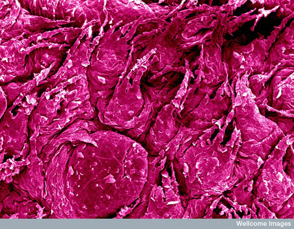
Surface Of Tongue Magnified
(Bamboo leaf for David Gregory&Debbie Marshall, Wellcome Images)
“Scanning electron microscopes image of the surface of the tongue, computer-colored: red/pink.”
Developing Sperm Under Microscope
")
(Bamboo leaf for Yorgos Nikas, Wellcome Images)
“Sperm develop in the seminiferous tubules of the testis. The spermatids are embedded in the Sertoli cells with their tails projecting into the lumen of the tubule. These sperm under the microscope are in the advanced stages of maturation. One of the spermatids has two tails (top right, green).”
Sperm Fertilizing An Egg Under A Microscope
")
(Bamboo leaf for Yorgos Nikas, Wellcome Images)
“Numerous sperm under a microscope trying to fertilize a human egg. They are trying to find their way through the zona pellucida, the membrane that surrounds and protects the egg.”
Close-Up Of Midge Eye
")
(Bamboo leaf for David Gregory&Debbie Marshall, Wellcome Images)
Part Of A Midge Head Magnified
")
(Bamboo leaf for David Gregory&Debbie Marshall, Wellcome Images)
Electron Microscope Image Of A Pubic Louse
")
Head of Pubic Louse
")
Claws of Pubic Louse
")
(Bamboo leaf for David Gregory&Debbie Marshall, Wellcome Images)
Stinging Hairs On A Nettle Leaf
")
(Bamboo leaf for David Liz Hirst, Wellcome Images)
“The large stinging hairs are hollow tubes with walls of silica making them into tiny glass needles. The bulb at the base of each hair contains the stinging liquid that includes formic acid, histamine, acetylcholine and 5- hydroxytryptamine (serotonin). The tips of the glassy hairs are very easily broken when brushed, leaving a sharp point, which easily pierces the skin to deliver the sting.”
Comparing Human Hair With Scanning Electron Microscopes
")
(Bamboo leaf for Anne Weston, Wellcome Images)
“This electron microscope image shows the difference in thickness of human hair between different ethnic groups. The strand of hair on the left is that of a Caucasian blond female. The hair on the right is from an Asian male. Hair is normally comprised of three layers, the inner medulla, the cortex, and the cuticle. The cuticle is the outermost layer and is comprised of numerous overlapping cells or scales. The cortex makes up the majority of the hair thickness. Interestingly, the inner medulla is not present in blond hair.”
Electron Microscope Image Of Human Tooth
")
(Bamboo leaf for David Gregory&Debbie Marshall, Wellcome Images)
“Low power scanning electron microscope image of tooth surface, computer-colored: yellow on blue background.”
Nanowire Magnified
")
(Bamboo leaf for Limin Tong/Harvard University)
“Electron microscope image of a nanowire curled into a loop in front of a strand of human hair. Nanowires can be as slender as 50 nanometers in width, about one-thousandth the width of a hair.”
Nanowires could be used in the near future to link tiny components into extremely small circuits.
Electron Microscope View Of Shark Skin
")
Source: unknown
“A scanning electron microscope reveals a shark’s secret to speed: tooth-like scales called dermal denticles. Water “races through the microgrooves without tumbling,” says shark researcher George Burgess, reducing friction. “It’s like a fast-moving river current versus the gurgling turbulence of a shallow stream.” The scales also discourage barnacles and algae from glomming on — an inspiration for synthetic coatings that may soon be applied to Navy ship hulls to reduce such biofouling.” They also give the shark’s skin the feel of sandpaper.
GO TO PART I
183Kviews
Share on Facebook


2
9